Zinc »
PDB 2fos-2g2p »
2fps »
Zinc in PDB 2fps: Crystal Structure of the N-Terminal Domain of E.Coli Hisb- Apo Ca Model.
Enzymatic activity of Crystal Structure of the N-Terminal Domain of E.Coli Hisb- Apo Ca Model.
All present enzymatic activity of Crystal Structure of the N-Terminal Domain of E.Coli Hisb- Apo Ca Model.:
3.1.3.15;
3.1.3.15;
Protein crystallography data
The structure of Crystal Structure of the N-Terminal Domain of E.Coli Hisb- Apo Ca Model., PDB code: 2fps
was solved by
E.S.Rangarajan,
M.Cygler,
A.Matte,
Montreal-Kingston Bacterialstructural Genomics Initiative (Bsgi),
with X-Ray Crystallography technique. A brief refinement statistics is given in the table below:
| Resolution Low / High (Å) | 50.00 / 2.20 |
| Space group | C 2 2 21 |
| Cell size a, b, c (Å), α, β, γ (°) | 52.748, 131.325, 104.898, 90.00, 90.00, 90.00 |
| R / Rfree (%) | 17 / 24.4 |
Other elements in 2fps:
The structure of Crystal Structure of the N-Terminal Domain of E.Coli Hisb- Apo Ca Model. also contains other interesting chemical elements:
| Chlorine | (Cl) | 2 atoms |
| Calcium | (Ca) | 2 atoms |
Zinc Binding Sites:
The binding sites of Zinc atom in the Crystal Structure of the N-Terminal Domain of E.Coli Hisb- Apo Ca Model.
(pdb code 2fps). This binding sites where shown within
5.0 Angstroms radius around Zinc atom.
In total 2 binding sites of Zinc where determined in the Crystal Structure of the N-Terminal Domain of E.Coli Hisb- Apo Ca Model., PDB code: 2fps:
Jump to Zinc binding site number: 1; 2;
In total 2 binding sites of Zinc where determined in the Crystal Structure of the N-Terminal Domain of E.Coli Hisb- Apo Ca Model., PDB code: 2fps:
Jump to Zinc binding site number: 1; 2;
Zinc binding site 1 out of 2 in 2fps
Go back to
Zinc binding site 1 out
of 2 in the Crystal Structure of the N-Terminal Domain of E.Coli Hisb- Apo Ca Model.
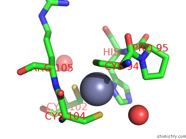
Mono view
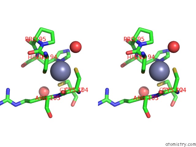
Stereo pair view
Mono view
Stereo pair view
A full contact list of Zinc with other atoms in the Zn binding
site number 1 of Crystal Structure of the N-Terminal Domain of E.Coli Hisb- Apo Ca Model. within 5.0Å range:
|
Zinc binding site 2 out of 2 in 2fps
Go back to
Zinc binding site 2 out
of 2 in the Crystal Structure of the N-Terminal Domain of E.Coli Hisb- Apo Ca Model.
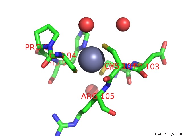
Mono view
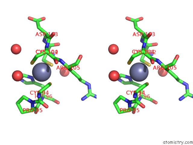
Stereo pair view
Mono view
Stereo pair view
A full contact list of Zinc with other atoms in the Zn binding
site number 2 of Crystal Structure of the N-Terminal Domain of E.Coli Hisb- Apo Ca Model. within 5.0Å range:
|
Reference:
E.S.Rangarajan,
A.Proteau,
J.Wagner,
M.N.Hung,
A.Matte,
M.Cygler.
Structural Snapshots of Escherichia Coli Histidinol Phosphate Phosphatase Along the Reaction Pathway. J.Biol.Chem. V. 281 37930 2006.
ISSN: ISSN 0021-9258
PubMed: 16966333
DOI: 10.1074/JBC.M604916200
Page generated: Wed Oct 16 23:51:31 2024
ISSN: ISSN 0021-9258
PubMed: 16966333
DOI: 10.1074/JBC.M604916200
Last articles
I in 1GZAI in 1GWD
I in 1GUL
I in 1GTE
I in 1GTH
I in 1GJD
I in 1F3M
I in 1FZ9
I in 1GA5
I in 1F86