Zinc »
PDB 2exf-2fa7 »
2f8b »
Zinc in PDB 2f8b: uc(Nmr) Structure of the C-Terminal Domain (Dimer) of HPV45 Oncoprotein E7
Zinc Binding Sites:
The binding sites of Zinc atom in the uc(Nmr) Structure of the C-Terminal Domain (Dimer) of HPV45 Oncoprotein E7
(pdb code 2f8b). This binding sites where shown within
5.0 Angstroms radius around Zinc atom.
In total 2 binding sites of Zinc where determined in the uc(Nmr) Structure of the C-Terminal Domain (Dimer) of HPV45 Oncoprotein E7, PDB code: 2f8b:
Jump to Zinc binding site number: 1; 2;
In total 2 binding sites of Zinc where determined in the uc(Nmr) Structure of the C-Terminal Domain (Dimer) of HPV45 Oncoprotein E7, PDB code: 2f8b:
Jump to Zinc binding site number: 1; 2;
Zinc binding site 1 out of 2 in 2f8b
Go back to
Zinc binding site 1 out
of 2 in the uc(Nmr) Structure of the C-Terminal Domain (Dimer) of HPV45 Oncoprotein E7
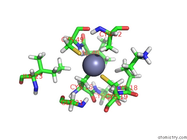
Mono view
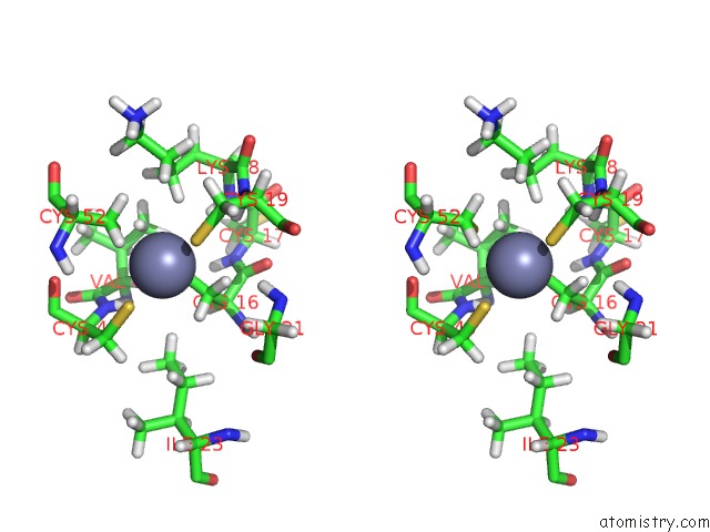
Stereo pair view
Mono view
Stereo pair view
A full contact list of Zinc with other atoms in the Zn binding
site number 1 of uc(Nmr) Structure of the C-Terminal Domain (Dimer) of HPV45 Oncoprotein E7 within 5.0Å range:
|
Zinc binding site 2 out of 2 in 2f8b
Go back to
Zinc binding site 2 out
of 2 in the uc(Nmr) Structure of the C-Terminal Domain (Dimer) of HPV45 Oncoprotein E7
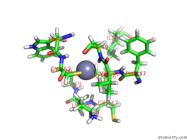
Mono view
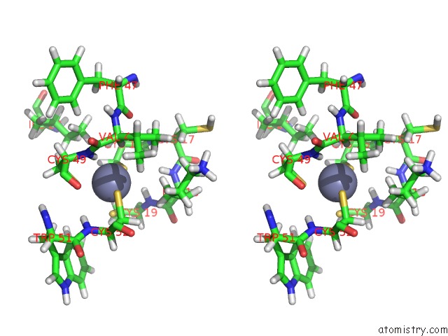
Stereo pair view
Mono view
Stereo pair view
A full contact list of Zinc with other atoms in the Zn binding
site number 2 of uc(Nmr) Structure of the C-Terminal Domain (Dimer) of HPV45 Oncoprotein E7 within 5.0Å range:
|
Reference:
O.Ohlenschlager,
T.Seiboth,
H.Zengerling,
L.Briese,
A.Marchanka,
R.Ramachandran,
M.Baum,
M.Korbas,
W.Meyer-Klaucke,
M.Durst,
M.Gorlach.
Solution Structure of the Partially Folded High-Risk Human Papilloma Virus 45 Oncoprotein E7. Oncogene V. 25 5953 2006.
ISSN: ISSN 0950-9232
PubMed: 16636661
DOI: 10.1038/SJ.ONC.1209584
Page generated: Wed Oct 16 23:40:45 2024
ISSN: ISSN 0950-9232
PubMed: 16636661
DOI: 10.1038/SJ.ONC.1209584
Last articles
Fe in 2YXOFe in 2YRS
Fe in 2YXC
Fe in 2YNM
Fe in 2YVJ
Fe in 2YP1
Fe in 2YU2
Fe in 2YU1
Fe in 2YQB
Fe in 2YOO