Zinc »
PDB 2eex-2elu »
2eg4 »
Zinc in PDB 2eg4: Crystal Structure of Probable Thiosulfate Sulfurtransferase
Protein crystallography data
The structure of Crystal Structure of Probable Thiosulfate Sulfurtransferase, PDB code: 2eg4
was solved by
H.Sakai,
A.Ebihara,
Y.Kitamura,
A.Shinkai,
S.Kuramitsu,
S.Yokoyama,
Rikenstructural Genomics/Proteomics Initiative (Rsgi),
with X-Ray Crystallography technique. A brief refinement statistics is given in the table below:
| Resolution Low / High (Å) | 32.81 / 1.70 |
| Space group | P 21 21 21 |
| Cell size a, b, c (Å), α, β, γ (°) | 62.694, 71.218, 115.483, 90.00, 90.00, 90.00 |
| R / Rfree (%) | 22.2 / 24.9 |
Zinc Binding Sites:
The binding sites of Zinc atom in the Crystal Structure of Probable Thiosulfate Sulfurtransferase
(pdb code 2eg4). This binding sites where shown within
5.0 Angstroms radius around Zinc atom.
In total 2 binding sites of Zinc where determined in the Crystal Structure of Probable Thiosulfate Sulfurtransferase, PDB code: 2eg4:
Jump to Zinc binding site number: 1; 2;
In total 2 binding sites of Zinc where determined in the Crystal Structure of Probable Thiosulfate Sulfurtransferase, PDB code: 2eg4:
Jump to Zinc binding site number: 1; 2;
Zinc binding site 1 out of 2 in 2eg4
Go back to
Zinc binding site 1 out
of 2 in the Crystal Structure of Probable Thiosulfate Sulfurtransferase
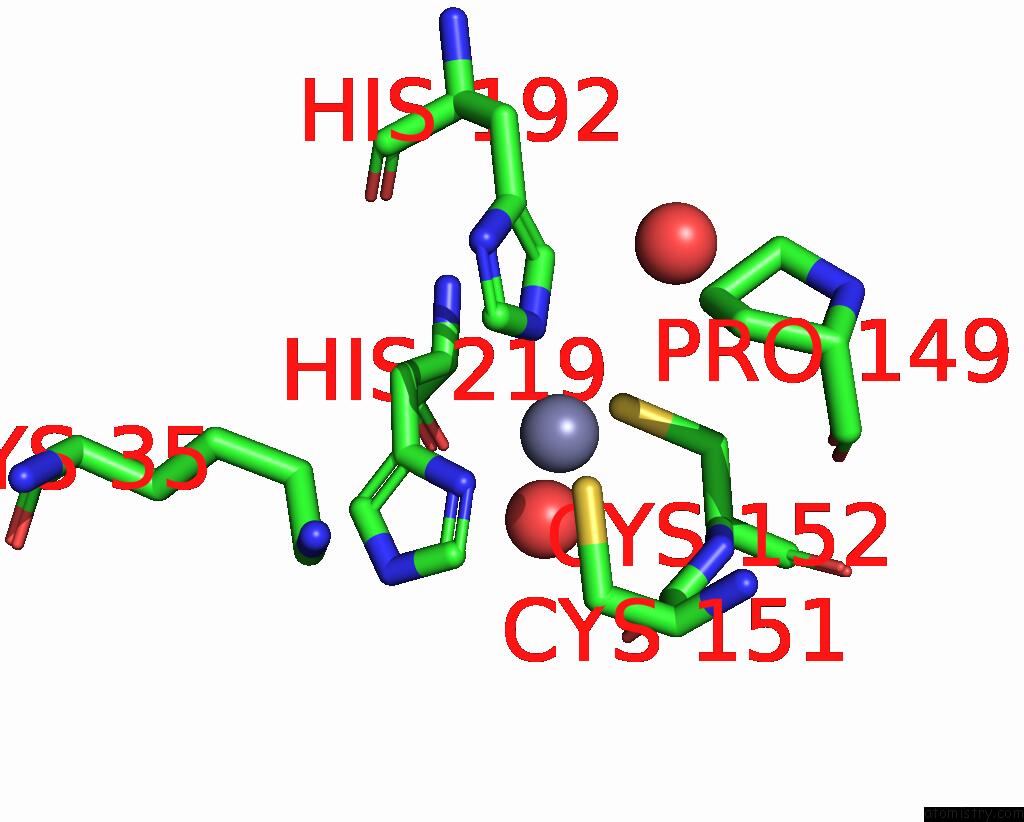
Mono view
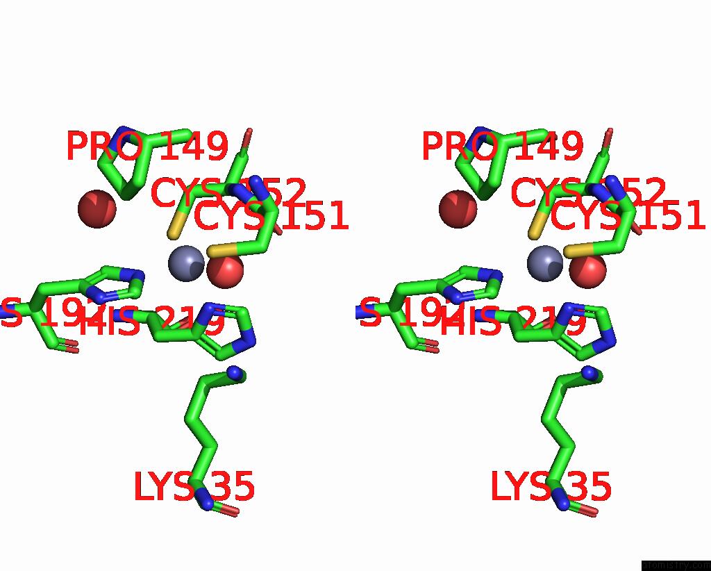
Stereo pair view
Mono view
Stereo pair view
A full contact list of Zinc with other atoms in the Zn binding
site number 1 of Crystal Structure of Probable Thiosulfate Sulfurtransferase within 5.0Å range:
|
Zinc binding site 2 out of 2 in 2eg4
Go back to
Zinc binding site 2 out
of 2 in the Crystal Structure of Probable Thiosulfate Sulfurtransferase
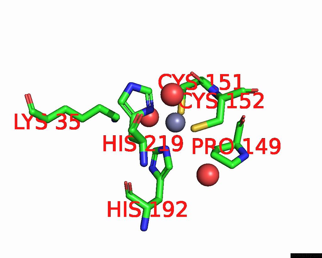
Mono view

Stereo pair view
Mono view
Stereo pair view
A full contact list of Zinc with other atoms in the Zn binding
site number 2 of Crystal Structure of Probable Thiosulfate Sulfurtransferase within 5.0Å range:
|
Reference:
H.Sakai,
A.Ebihara,
Y.Kitamura,
A.Shinkai,
S.Kuramitsu,
S.Yokoyama.
Crystal Structure of Probable Thiosulfate Sulfurtransferase To Be Published.
Page generated: Wed Oct 16 23:06:49 2024
Last articles
Mg in 5NG1Mg in 5N9Z
Mg in 5NH2
Mg in 5NFZ
Mg in 5NFV
Mg in 5NG0
Mg in 5NEU
Mg in 5NDH
Mg in 5ND7
Mg in 5ND4