Zinc »
PDB 2e3x-2eer »
2e3x »
Zinc in PDB 2e3x: Crystal Structure of Russell'S Viper Venom Metalloproteinase
Enzymatic activity of Crystal Structure of Russell'S Viper Venom Metalloproteinase
All present enzymatic activity of Crystal Structure of Russell'S Viper Venom Metalloproteinase:
3.4.24.58;
3.4.24.58;
Protein crystallography data
The structure of Crystal Structure of Russell'S Viper Venom Metalloproteinase, PDB code: 2e3x
was solved by
T.Igarashi,
S.Takeda,
with X-Ray Crystallography technique. A brief refinement statistics is given in the table below:
| Resolution Low / High (Å) | 44.60 / 2.91 |
| Space group | P 21 21 21 |
| Cell size a, b, c (Å), α, β, γ (°) | 70.350, 91.730, 152.930, 90.00, 90.00, 90.00 |
| R / Rfree (%) | 21.8 / 27.3 |
Other elements in 2e3x:
The structure of Crystal Structure of Russell'S Viper Venom Metalloproteinase also contains other interesting chemical elements:
| Calcium | (Ca) | 5 atoms |
Zinc Binding Sites:
The binding sites of Zinc atom in the Crystal Structure of Russell'S Viper Venom Metalloproteinase
(pdb code 2e3x). This binding sites where shown within
5.0 Angstroms radius around Zinc atom.
In total only one binding site of Zinc was determined in the Crystal Structure of Russell'S Viper Venom Metalloproteinase, PDB code: 2e3x:
In total only one binding site of Zinc was determined in the Crystal Structure of Russell'S Viper Venom Metalloproteinase, PDB code: 2e3x:
Zinc binding site 1 out of 1 in 2e3x
Go back to
Zinc binding site 1 out
of 1 in the Crystal Structure of Russell'S Viper Venom Metalloproteinase
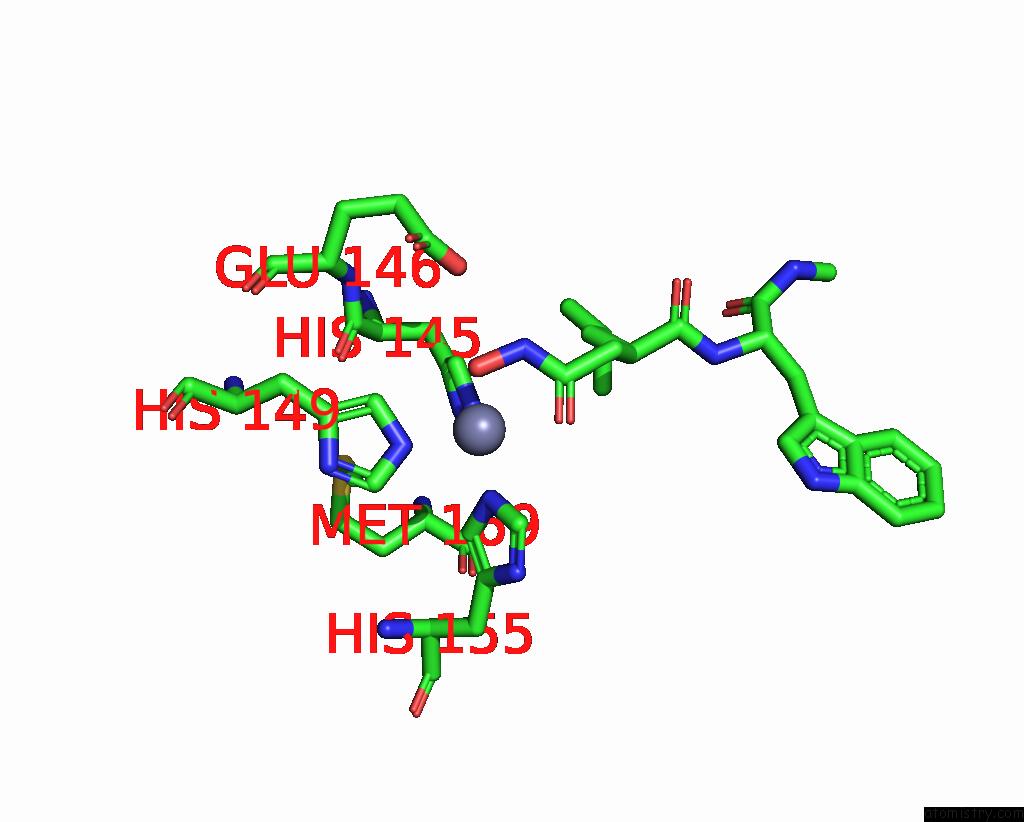
Mono view
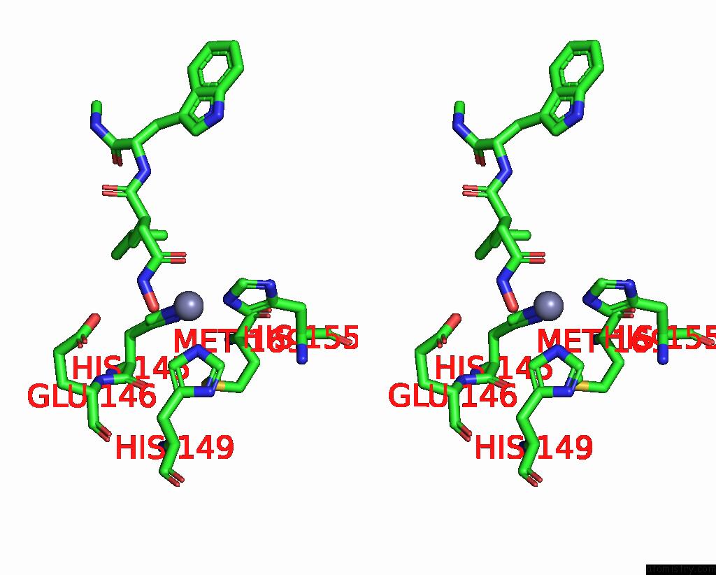
Stereo pair view
Mono view
Stereo pair view
A full contact list of Zinc with other atoms in the Zn binding
site number 1 of Crystal Structure of Russell'S Viper Venom Metalloproteinase within 5.0Å range:
|
Reference:
S.Takeda,
T.Igarashi,
H.Mori.
Crystal Structure of Rvv-X: An Example of Evolutionary Gain of Specificity By Adam Proteinases. Febs Lett. V. 581 5859 2007.
ISSN: ISSN 0014-5793
PubMed: 18060879
DOI: 10.1016/J.FEBSLET.2007.11.062
Page generated: Wed Oct 16 22:58:30 2024
ISSN: ISSN 0014-5793
PubMed: 18060879
DOI: 10.1016/J.FEBSLET.2007.11.062
Last articles
Fe in 2YXOFe in 2YRS
Fe in 2YXC
Fe in 2YNM
Fe in 2YVJ
Fe in 2YP1
Fe in 2YU2
Fe in 2YU1
Fe in 2YQB
Fe in 2YOO