Zinc »
PDB 2cim-2ctd »
2cs7 »
Zinc in PDB 2cs7: 1.2 A Crystal Structure of the S. Pneumoniae Phta Histidine Triad Domain A Novel Zinc Binding Fold
Protein crystallography data
The structure of 1.2 A Crystal Structure of the S. Pneumoniae Phta Histidine Triad Domain A Novel Zinc Binding Fold, PDB code: 2cs7
was solved by
A.Riboldi-Tunnicliffe,
N.W.Isaacs,
T.J.Mitchell,
with X-Ray Crystallography technique. A brief refinement statistics is given in the table below:
| Resolution Low / High (Å) | 72.55 / 1.20 |
| Space group | C 1 2 1 |
| Cell size a, b, c (Å), α, β, γ (°) | 62.182, 35.895, 72.541, 90.00, 90.01, 90.00 |
| R / Rfree (%) | 11.2 / 13.5 |
Zinc Binding Sites:
The binding sites of Zinc atom in the 1.2 A Crystal Structure of the S. Pneumoniae Phta Histidine Triad Domain A Novel Zinc Binding Fold
(pdb code 2cs7). This binding sites where shown within
5.0 Angstroms radius around Zinc atom.
In total 3 binding sites of Zinc where determined in the 1.2 A Crystal Structure of the S. Pneumoniae Phta Histidine Triad Domain A Novel Zinc Binding Fold, PDB code: 2cs7:
Jump to Zinc binding site number: 1; 2; 3;
In total 3 binding sites of Zinc where determined in the 1.2 A Crystal Structure of the S. Pneumoniae Phta Histidine Triad Domain A Novel Zinc Binding Fold, PDB code: 2cs7:
Jump to Zinc binding site number: 1; 2; 3;
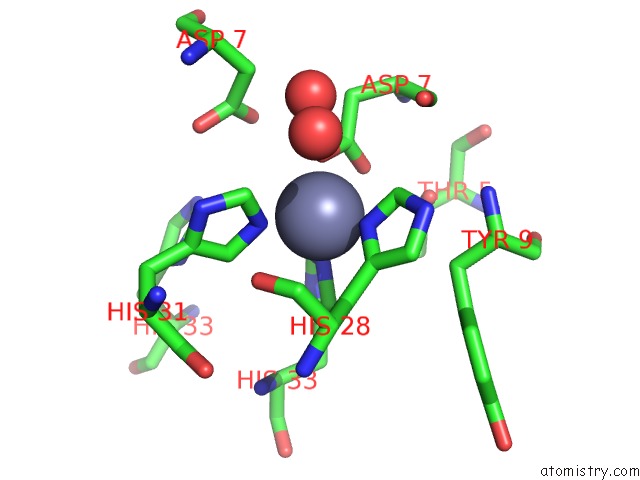
Zinc binding site 1 out of 3 in 2cs7
Go back to
Zinc binding site 1 out
of 3 in the 1.2 A Crystal Structure of the S. Pneumoniae Phta Histidine Triad Domain A Novel Zinc Binding Fold
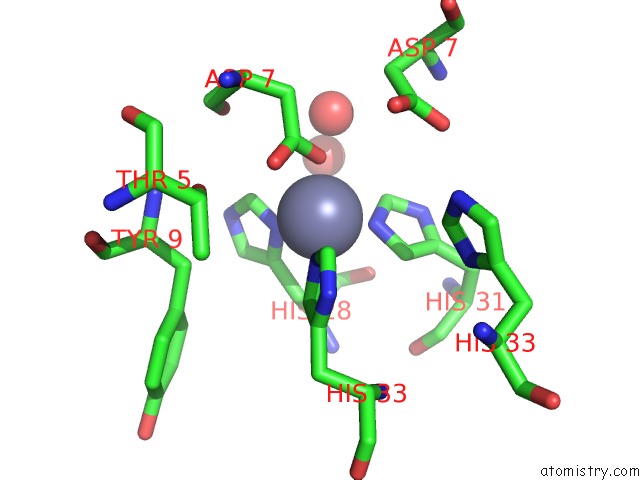
Mono view
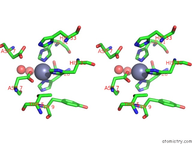
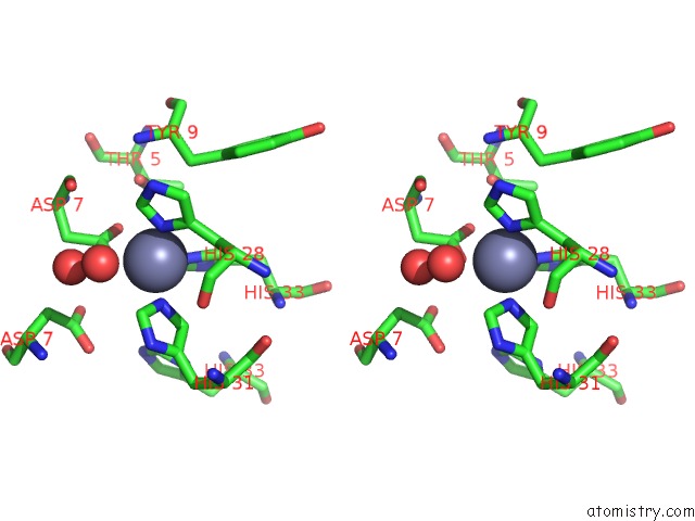
Stereo pair view
Mono view
Stereo pair view
A full contact list of Zinc with other atoms in the Zn binding
site number 1 of 1.2 A Crystal Structure of the S. Pneumoniae Phta Histidine Triad Domain A Novel Zinc Binding Fold within 5.0Å range:
|
Zinc binding site 2 out of 3 in 2cs7
Go back to
Zinc binding site 2 out
of 3 in the 1.2 A Crystal Structure of the S. Pneumoniae Phta Histidine Triad Domain A Novel Zinc Binding Fold
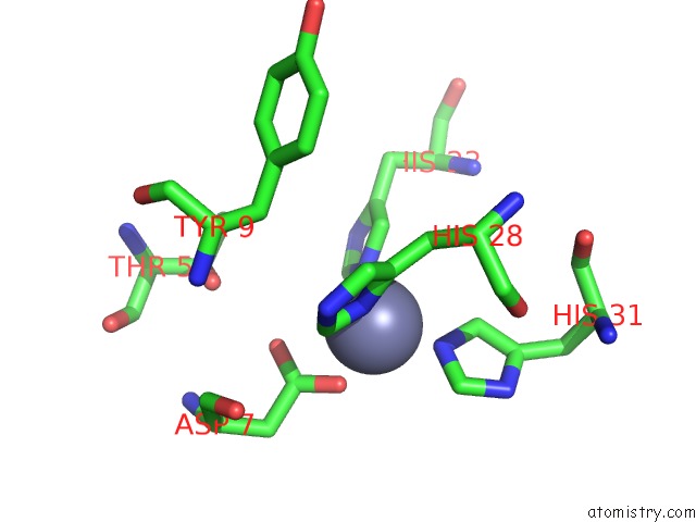
Mono view
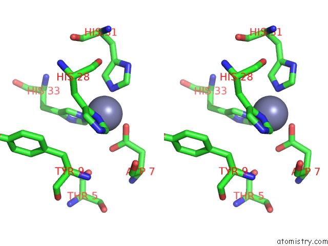
Stereo pair view
Mono view
Stereo pair view
A full contact list of Zinc with other atoms in the Zn binding
site number 2 of 1.2 A Crystal Structure of the S. Pneumoniae Phta Histidine Triad Domain A Novel Zinc Binding Fold within 5.0Å range:
|
Zinc binding site 3 out of 3 in 2cs7
Go back to
Zinc binding site 3 out
of 3 in the 1.2 A Crystal Structure of the S. Pneumoniae Phta Histidine Triad Domain A Novel Zinc Binding Fold
Mono view
Stereo pair view
Mono view
Stereo pair view
A full contact list of Zinc with other atoms in the Zn binding
site number 3 of 1.2 A Crystal Structure of the S. Pneumoniae Phta Histidine Triad Domain A Novel Zinc Binding Fold within 5.0Å range:
|
Reference:
A.Riboldi-Tunnicliffe,
N.W.Isaacs,
T.J.Mitchell.
1.2 Angstroms Crystal Structure of the S. Pneumoniae Phta Histidine Triad Domain A Novel Zinc Binding Fold. Febs Lett. V. 579 5353 2005.
ISSN: ISSN 0014-5793
PubMed: 16194532
DOI: 10.1016/J.FEBSLET.2005.08.066
Page generated: Wed Oct 16 22:29:47 2024
ISSN: ISSN 0014-5793
PubMed: 16194532
DOI: 10.1016/J.FEBSLET.2005.08.066
Last articles
Mn in 4UXAMn in 4W8Y
Mn in 4W9S
Mn in 4V15
Mn in 4V0U
Mn in 4V0W
Mn in 4V0X
Mn in 4UWQ
Mn in 4V0V
Mn in 4URR