Zinc »
PDB 2a2q-2afm »
2ac3 »
Zinc in PDB 2ac3: Structure of Human MNK2 Kinase Domain
Enzymatic activity of Structure of Human MNK2 Kinase Domain
All present enzymatic activity of Structure of Human MNK2 Kinase Domain:
2.7.1.37;
2.7.1.37;
Protein crystallography data
The structure of Structure of Human MNK2 Kinase Domain, PDB code: 2ac3
was solved by
R.Jauch,
M.C.Wahl,
C.Netter,
S.Jakel,
K.Schreiter,
B.Aicher,
H.Jackle,
with X-Ray Crystallography technique. A brief refinement statistics is given in the table below:
| Resolution Low / High (Å) | 15.00 / 2.10 |
| Space group | P 32 2 1 |
| Cell size a, b, c (Å), α, β, γ (°) | 104.502, 104.502, 72.351, 90.00, 90.00, 120.00 |
| R / Rfree (%) | 21.5 / 25.4 |
Zinc Binding Sites:
The binding sites of Zinc atom in the Structure of Human MNK2 Kinase Domain
(pdb code 2ac3). This binding sites where shown within
5.0 Angstroms radius around Zinc atom.
In total only one binding site of Zinc was determined in the Structure of Human MNK2 Kinase Domain, PDB code: 2ac3:
In total only one binding site of Zinc was determined in the Structure of Human MNK2 Kinase Domain, PDB code: 2ac3:
Zinc binding site 1 out of 1 in 2ac3
Go back to
Zinc binding site 1 out
of 1 in the Structure of Human MNK2 Kinase Domain
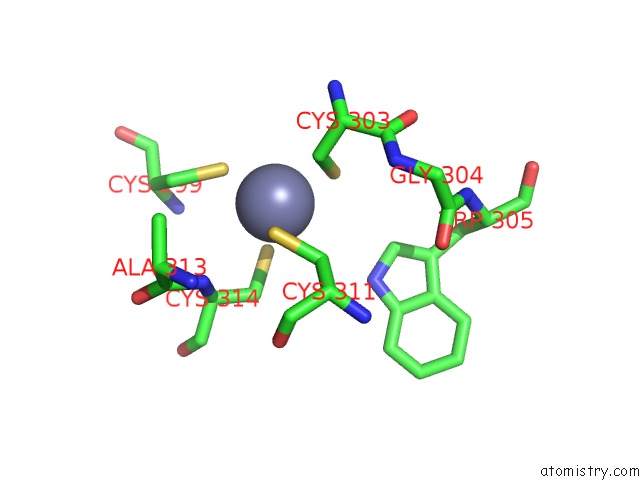
Mono view
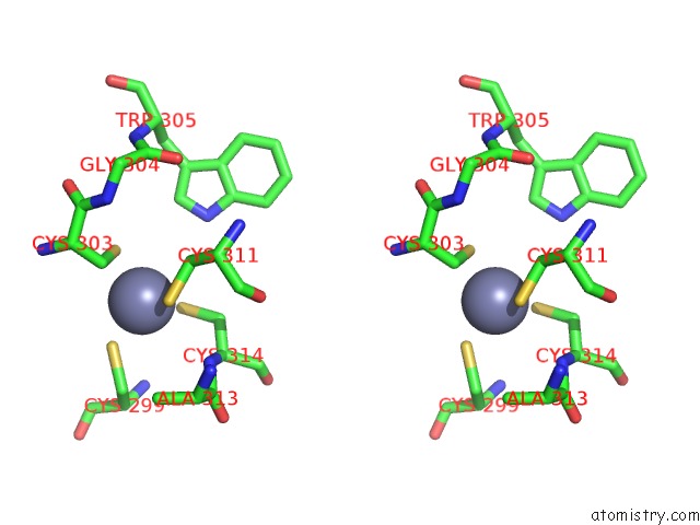
Stereo pair view
Mono view
Stereo pair view
A full contact list of Zinc with other atoms in the Zn binding
site number 1 of Structure of Human MNK2 Kinase Domain within 5.0Å range:
|
Reference:
R.Jauch,
S.Jakel,
C.Netter,
K.Schreiter,
B.Aicher,
H.Jackle,
M.C.Wahl.
Crystal Structures of the MNK2 Kinase Domain Reveal An Inhibitory Conformation and A Zinc Binding Site. Structure V. 13 1559 2005.
ISSN: ISSN 0969-2126
PubMed: 16216586
DOI: 10.1016/J.STR.2005.07.013
Page generated: Wed Oct 16 21:35:54 2024
ISSN: ISSN 0969-2126
PubMed: 16216586
DOI: 10.1016/J.STR.2005.07.013
Last articles
Mg in 6E7CMg in 6E7B
Mg in 6E6P
Mg in 6E6H
Mg in 6E6F
Mg in 6E61
Mg in 6E60
Mg in 6E6C
Mg in 6E53
Mg in 6E3X