Zinc »
PDB 1ylk-1z5h »
1yp1 »
Zinc in PDB 1yp1: Crystal Structure of A Non-Hemorrhagic Fibrin(Ogen)Olytic Metalloproteinase From Venom of Agkistrodon Acutus
Protein crystallography data
The structure of Crystal Structure of A Non-Hemorrhagic Fibrin(Ogen)Olytic Metalloproteinase From Venom of Agkistrodon Acutus, PDB code: 1yp1
was solved by
Z.Lou,
J.Hou,
J.Chen,
X.Liang,
P.Qiu,
Y.Liu,
M.Li,
Z.Rao,
with X-Ray Crystallography technique. A brief refinement statistics is given in the table below:
| Resolution Low / High (Å) | 50.00 / 1.90 |
| Space group | P 31 2 1 |
| Cell size a, b, c (Å), α, β, γ (°) | 80.565, 80.565, 66.769, 90.00, 90.00, 120.00 |
| R / Rfree (%) | 21.7 / 23.4 |
Zinc Binding Sites:
The binding sites of Zinc atom in the Crystal Structure of A Non-Hemorrhagic Fibrin(Ogen)Olytic Metalloproteinase From Venom of Agkistrodon Acutus
(pdb code 1yp1). This binding sites where shown within
5.0 Angstroms radius around Zinc atom.
In total only one binding site of Zinc was determined in the Crystal Structure of A Non-Hemorrhagic Fibrin(Ogen)Olytic Metalloproteinase From Venom of Agkistrodon Acutus, PDB code: 1yp1:
In total only one binding site of Zinc was determined in the Crystal Structure of A Non-Hemorrhagic Fibrin(Ogen)Olytic Metalloproteinase From Venom of Agkistrodon Acutus, PDB code: 1yp1:
Zinc binding site 1 out of 1 in 1yp1
Go back to
Zinc binding site 1 out
of 1 in the Crystal Structure of A Non-Hemorrhagic Fibrin(Ogen)Olytic Metalloproteinase From Venom of Agkistrodon Acutus
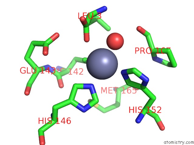
Mono view
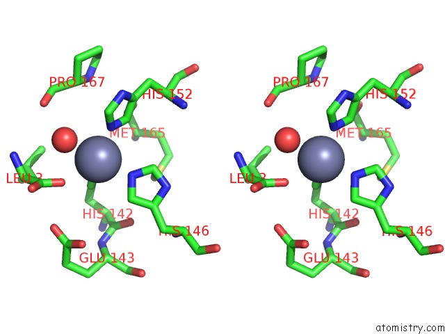
Stereo pair view
Mono view
Stereo pair view
A full contact list of Zinc with other atoms in the Zn binding
site number 1 of Crystal Structure of A Non-Hemorrhagic Fibrin(Ogen)Olytic Metalloproteinase From Venom of Agkistrodon Acutus within 5.0Å range:
|
Reference:
Z.Lou,
J.Hou,
X.Liang,
J.Chen,
P.Qiu,
Y.Liu,
M.Li,
Z.Rao,
G.Yan.
Crystal Structure of A Non-Hemorrhagic Fibrin(Ogen)Olytic Metalloproteinase Complexed with A Novel Natural Tri-Peptide Inhibitor From Venom of Agkistrodon Acutus J.Struct.Biol. V. 152 195 2005.
ISSN: ISSN 1047-8477
PubMed: 16330227
DOI: 10.1016/J.JSB.2005.09.006
Page generated: Wed Oct 16 21:02:23 2024
ISSN: ISSN 1047-8477
PubMed: 16330227
DOI: 10.1016/J.JSB.2005.09.006
Last articles
Mg in 3CCQMg in 3CCM
Mg in 3CCL
Mg in 3CCJ
Mg in 3CC2
Mg in 3CCE
Mg in 3CC7
Mg in 3CC4
Mg in 3CB3
Mg in 3CC6