Zinc »
PDB 1ylk-1z5h »
1ynw »
Zinc in PDB 1ynw: Crystal Structure of Vitamin D Receptor and 9-Cis Retinoic Acid Receptor Dna-Binding Domains Bound to A DR3 Response Element
Protein crystallography data
The structure of Crystal Structure of Vitamin D Receptor and 9-Cis Retinoic Acid Receptor Dna-Binding Domains Bound to A DR3 Response Element, PDB code: 1ynw
was solved by
P.L.Shaffer,
D.T.Gewirth,
with X-Ray Crystallography technique. A brief refinement statistics is given in the table below:
| Resolution Low / High (Å) | 50.00 / 3.00 |
| Space group | C 1 2 1 |
| Cell size a, b, c (Å), α, β, γ (°) | 123.104, 57.049, 73.438, 90.00, 110.31, 90.00 |
| R / Rfree (%) | 23.2 / 28.3 |
Zinc Binding Sites:
The binding sites of Zinc atom in the Crystal Structure of Vitamin D Receptor and 9-Cis Retinoic Acid Receptor Dna-Binding Domains Bound to A DR3 Response Element
(pdb code 1ynw). This binding sites where shown within
5.0 Angstroms radius around Zinc atom.
In total 4 binding sites of Zinc where determined in the Crystal Structure of Vitamin D Receptor and 9-Cis Retinoic Acid Receptor Dna-Binding Domains Bound to A DR3 Response Element, PDB code: 1ynw:
Jump to Zinc binding site number: 1; 2; 3; 4;
In total 4 binding sites of Zinc where determined in the Crystal Structure of Vitamin D Receptor and 9-Cis Retinoic Acid Receptor Dna-Binding Domains Bound to A DR3 Response Element, PDB code: 1ynw:
Jump to Zinc binding site number: 1; 2; 3; 4;
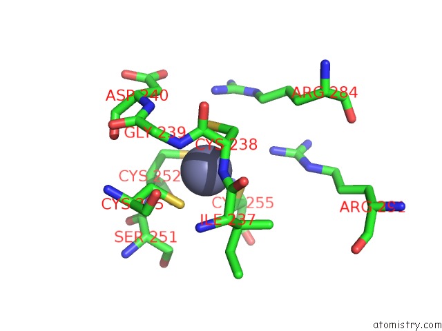
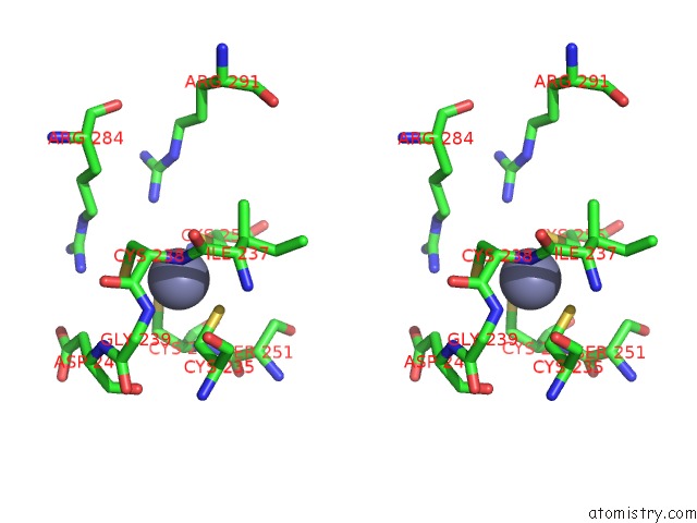
Zinc binding site 1 out of 4 in 1ynw
Go back to
Zinc binding site 1 out
of 4 in the Crystal Structure of Vitamin D Receptor and 9-Cis Retinoic Acid Receptor Dna-Binding Domains Bound to A DR3 Response Element
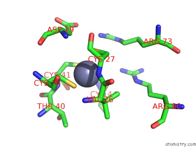
Mono view
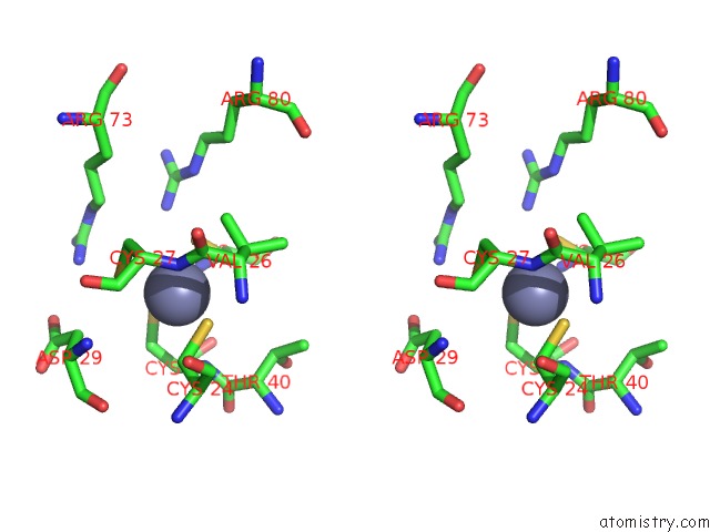
Stereo pair view
Mono view
Stereo pair view
A full contact list of Zinc with other atoms in the Zn binding
site number 1 of Crystal Structure of Vitamin D Receptor and 9-Cis Retinoic Acid Receptor Dna-Binding Domains Bound to A DR3 Response Element within 5.0Å range:
|
Zinc binding site 2 out of 4 in 1ynw
Go back to
Zinc binding site 2 out
of 4 in the Crystal Structure of Vitamin D Receptor and 9-Cis Retinoic Acid Receptor Dna-Binding Domains Bound to A DR3 Response Element
Mono view
Stereo pair view
Mono view
Stereo pair view
A full contact list of Zinc with other atoms in the Zn binding
site number 2 of Crystal Structure of Vitamin D Receptor and 9-Cis Retinoic Acid Receptor Dna-Binding Domains Bound to A DR3 Response Element within 5.0Å range:
|
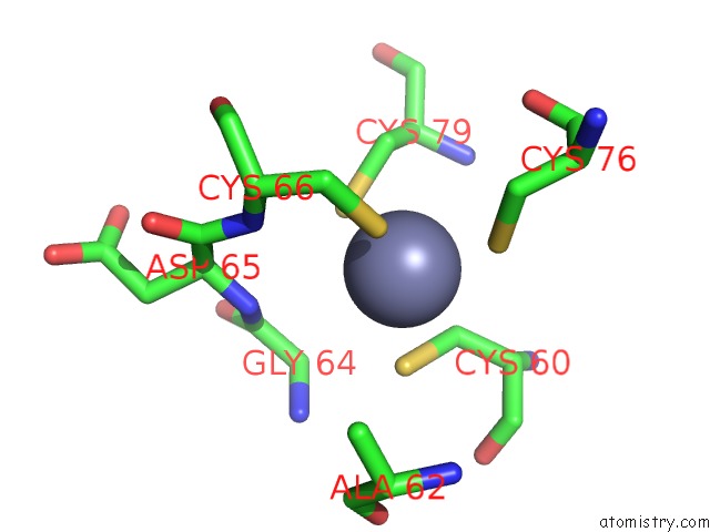
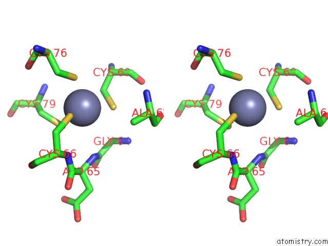
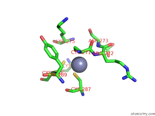
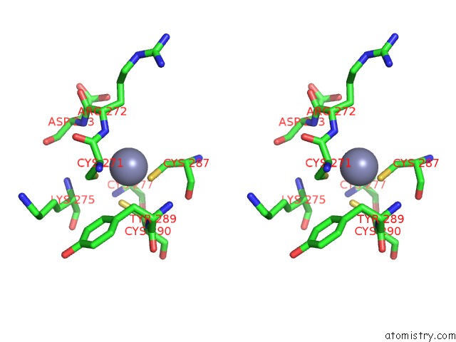
Zinc binding site 3 out of 4 in 1ynw
Go back to
Zinc binding site 3 out
of 4 in the Crystal Structure of Vitamin D Receptor and 9-Cis Retinoic Acid Receptor Dna-Binding Domains Bound to A DR3 Response Element
Mono view
Stereo pair view
Mono view
Stereo pair view
A full contact list of Zinc with other atoms in the Zn binding
site number 3 of Crystal Structure of Vitamin D Receptor and 9-Cis Retinoic Acid Receptor Dna-Binding Domains Bound to A DR3 Response Element within 5.0Å range:
|
Zinc binding site 4 out of 4 in 1ynw
Go back to
Zinc binding site 4 out
of 4 in the Crystal Structure of Vitamin D Receptor and 9-Cis Retinoic Acid Receptor Dna-Binding Domains Bound to A DR3 Response Element
Mono view
Stereo pair view
Mono view
Stereo pair view
A full contact list of Zinc with other atoms in the Zn binding
site number 4 of Crystal Structure of Vitamin D Receptor and 9-Cis Retinoic Acid Receptor Dna-Binding Domains Bound to A DR3 Response Element within 5.0Å range:
|
Reference:
P.L.Shaffer,
D.T.Gewirth.
Structural Analysis of Rxr-Vdr Interactions on DR3 Dna J.Steroid Biochem.Mol.Biol. V.9-90 215 2004.
ISSN: ISSN 0960-0760
PubMed: 15225774
DOI: 10.1016/J.JSBMB.2004.03.084
Page generated: Wed Oct 16 21:00:43 2024
ISSN: ISSN 0960-0760
PubMed: 15225774
DOI: 10.1016/J.JSBMB.2004.03.084
Last articles
Mg in 3CCQMg in 3CCM
Mg in 3CCL
Mg in 3CCJ
Mg in 3CC2
Mg in 3CCE
Mg in 3CC7
Mg in 3CC4
Mg in 3CB3
Mg in 3CC6