Zinc »
PDB 1xv2-1y8j »
1y3g »
Zinc in PDB 1y3g: Crystal Structure of A Silanediol Protease Inhibitor Bound to Thermolysin
Enzymatic activity of Crystal Structure of A Silanediol Protease Inhibitor Bound to Thermolysin
All present enzymatic activity of Crystal Structure of A Silanediol Protease Inhibitor Bound to Thermolysin:
3.4.24.27;
3.4.24.27;
Protein crystallography data
The structure of Crystal Structure of A Silanediol Protease Inhibitor Bound to Thermolysin, PDB code: 1y3g
was solved by
D.H.Juers,
J.Kim,
B.W.Matthews,
S.M.Sieburth,
with X-Ray Crystallography technique. A brief refinement statistics is given in the table below:
| Resolution Low / High (Å) | 31.00 / 2.10 |
| Space group | P 61 2 2 |
| Cell size a, b, c (Å), α, β, γ (°) | 93.520, 93.520, 131.770, 90.00, 90.00, 120.00 |
| R / Rfree (%) | 15.5 / 22.5 |
Other elements in 1y3g:
The structure of Crystal Structure of A Silanediol Protease Inhibitor Bound to Thermolysin also contains other interesting chemical elements:
| Silicon | (Si) | 1 atom |
| Calcium | (Ca) | 4 atoms |
Zinc Binding Sites:
The binding sites of Zinc atom in the Crystal Structure of A Silanediol Protease Inhibitor Bound to Thermolysin
(pdb code 1y3g). This binding sites where shown within
5.0 Angstroms radius around Zinc atom.
In total only one binding site of Zinc was determined in the Crystal Structure of A Silanediol Protease Inhibitor Bound to Thermolysin, PDB code: 1y3g:
In total only one binding site of Zinc was determined in the Crystal Structure of A Silanediol Protease Inhibitor Bound to Thermolysin, PDB code: 1y3g:
Zinc binding site 1 out of 1 in 1y3g
Go back to
Zinc binding site 1 out
of 1 in the Crystal Structure of A Silanediol Protease Inhibitor Bound to Thermolysin
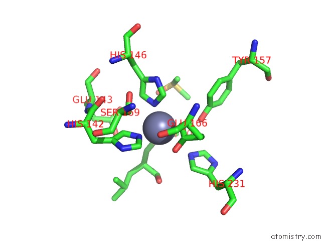
Mono view
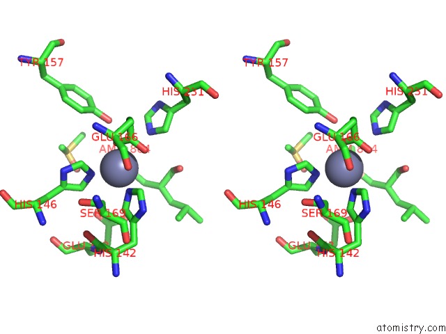
Stereo pair view
Mono view
Stereo pair view
A full contact list of Zinc with other atoms in the Zn binding
site number 1 of Crystal Structure of A Silanediol Protease Inhibitor Bound to Thermolysin within 5.0Å range:
|
Reference:
D.H.Juers,
J.Kim,
B.W.Matthews,
S.M.Sieburth.
Structural Analysis of Silanediols As Transition-State-Analogue Inhibitors of the Benchmark Metalloprotease Thermolysin(,). Biochemistry V. 44 16524 2005.
ISSN: ISSN 0006-2960
PubMed: 16342943
DOI: 10.1021/BI051346V
Page generated: Wed Oct 16 20:45:31 2024
ISSN: ISSN 0006-2960
PubMed: 16342943
DOI: 10.1021/BI051346V
Last articles
Mo in 6GAXMo in 6CZA
Mo in 6CZ9
Mo in 6CZ8
Mo in 6CZ7
Mo in 6BBL
Mo in 6CDK
Mo in 5WA0
Mo in 6A71
Mo in 6A72