Zinc »
PDB 1x8h-1xm8 »
1xjo »
Zinc in PDB 1xjo: Structure of Aminopeptidase
Protein crystallography data
The structure of Structure of Aminopeptidase, PDB code: 1xjo
was solved by
H.M.Greenblatt,
D.Barra,
S.Blumberg,
G.Shoham,
with X-Ray Crystallography technique. A brief refinement statistics is given in the table below:
| Resolution Low / High (Å) | 20.00 / 1.75 |
| Space group | P 41 21 2 |
| Cell size a, b, c (Å), α, β, γ (°) | 61.810, 61.810, 146.300, 90.00, 90.00, 90.00 |
| R / Rfree (%) | 14.1 / n/a |
Other elements in 1xjo:
The structure of Structure of Aminopeptidase also contains other interesting chemical elements:
| Calcium | (Ca) | 1 atom |
Zinc Binding Sites:
The binding sites of Zinc atom in the Structure of Aminopeptidase
(pdb code 1xjo). This binding sites where shown within
5.0 Angstroms radius around Zinc atom.
In total 2 binding sites of Zinc where determined in the Structure of Aminopeptidase, PDB code: 1xjo:
Jump to Zinc binding site number: 1; 2;
In total 2 binding sites of Zinc where determined in the Structure of Aminopeptidase, PDB code: 1xjo:
Jump to Zinc binding site number: 1; 2;
Zinc binding site 1 out of 2 in 1xjo
Go back to
Zinc binding site 1 out
of 2 in the Structure of Aminopeptidase
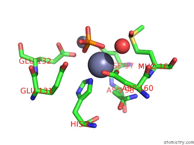
Mono view
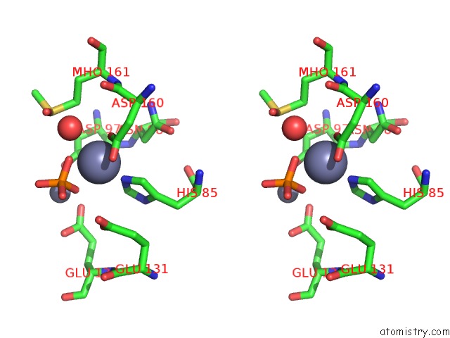
Stereo pair view
Mono view
Stereo pair view
A full contact list of Zinc with other atoms in the Zn binding
site number 1 of Structure of Aminopeptidase within 5.0Å range:
|
Zinc binding site 2 out of 2 in 1xjo
Go back to
Zinc binding site 2 out
of 2 in the Structure of Aminopeptidase
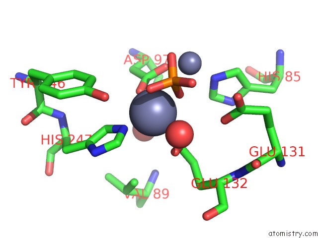
Mono view
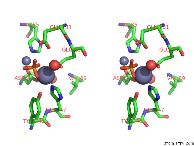
Stereo pair view
Mono view
Stereo pair view
A full contact list of Zinc with other atoms in the Zn binding
site number 2 of Structure of Aminopeptidase within 5.0Å range:
|
Reference:
H.M.Greenblatt,
O.Almog,
B.Maras,
A.Spungin-Bialik,
D.Barra,
S.Blumberg,
G.Shoham.
Streptomyces Griseus Aminopeptidase: X-Ray Crystallographic Structure at 1.75 A Resolution. J.Mol.Biol. V. 265 620 1997.
ISSN: ISSN 0022-2836
PubMed: 9048953
DOI: 10.1006/JMBI.1996.0729
Page generated: Wed Oct 16 20:28:16 2024
ISSN: ISSN 0022-2836
PubMed: 9048953
DOI: 10.1006/JMBI.1996.0729
Last articles
Mn in 5WE7Mn in 5WE9
Mn in 5WDW
Mn in 5WCX
Mn in 5WDN
Mn in 5WDC
Mn in 5WCT
Mn in 5WCS
Mn in 5WB3
Mn in 5WAP