Zinc »
PDB 1wfz-1wwd »
1wni »
Zinc in PDB 1wni: Crystal Structure of H2-Proteinase
Enzymatic activity of Crystal Structure of H2-Proteinase
All present enzymatic activity of Crystal Structure of H2-Proteinase:
3.4.24.53;
3.4.24.53;
Protein crystallography data
The structure of Crystal Structure of H2-Proteinase, PDB code: 1wni
was solved by
T.Kumasaka,
M.Yamamoto,
H.Moriyama,
N.Tanaka,
M.Sato,
Y.Katsube,
Y.Yamakawa,
T.Omori-Satoh,
S.Iwanaga,
T.Ueki,
with X-Ray Crystallography technique. A brief refinement statistics is given in the table below:
| Resolution Low / High (Å) | 8.00 / 2.20 |
| Space group | P 43 21 2 |
| Cell size a, b, c (Å), α, β, γ (°) | 77.800, 77.800, 82.300, 90.00, 90.00, 90.00 |
| R / Rfree (%) | 17.6 / n/a |
Zinc Binding Sites:
The binding sites of Zinc atom in the Crystal Structure of H2-Proteinase
(pdb code 1wni). This binding sites where shown within
5.0 Angstroms radius around Zinc atom.
In total only one binding site of Zinc was determined in the Crystal Structure of H2-Proteinase, PDB code: 1wni:
In total only one binding site of Zinc was determined in the Crystal Structure of H2-Proteinase, PDB code: 1wni:
Zinc binding site 1 out of 1 in 1wni
Go back to
Zinc binding site 1 out
of 1 in the Crystal Structure of H2-Proteinase
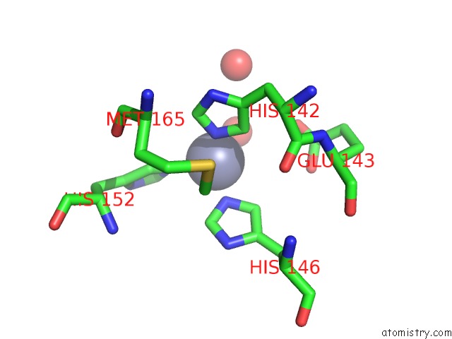
Mono view
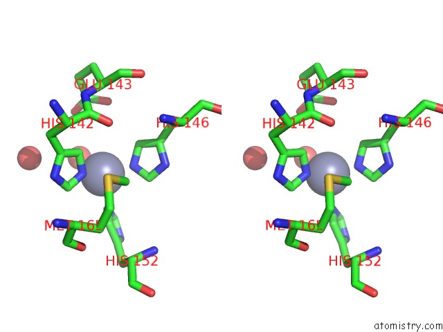
Stereo pair view
Mono view
Stereo pair view
A full contact list of Zinc with other atoms in the Zn binding
site number 1 of Crystal Structure of H2-Proteinase within 5.0Å range:
|
Reference:
T.Kumasaka,
M.Yamamoto,
H.Moriyama,
N.Tanaka,
M.Sato,
Y.Katsube,
Y.Yamakawa,
T.Omori-Satoh,
S.Iwanaga,
T.Ueki.
Crystal Structure of H2-Proteinase From the Venom of Trimeresurus Flavoviridis. J.Biochem. V. 119 49 1996.
ISSN: ISSN 0021-924X
PubMed: 8907175
DOI: 10.1093/OXFORDJOURNALS.JBCHEM.A021215
Page generated: Wed Oct 16 20:07:26 2024
ISSN: ISSN 0021-924X
PubMed: 8907175
DOI: 10.1093/OXFORDJOURNALS.JBCHEM.A021215
Last articles
Fe in 6MYPFe in 6MYO
Fe in 6MSN
Fe in 6MX5
Fe in 6MV0
Fe in 6MEV
Fe in 6MQ6
Fe in 6MQ1
Fe in 6MQ0
Fe in 6MPY