Zinc »
PDB 1vev-1w57 »
1vzy »
Zinc in PDB 1vzy: Crystal Structure of the Bacillus Subtilis HSP33
Protein crystallography data
The structure of Crystal Structure of the Bacillus Subtilis HSP33, PDB code: 1vzy
was solved by
I.K.Janda,
Y.Devedjiev,
U.Derewenda,
Z.Dauter,
J.Bielnicki,
D.R.Cooper,
A.Joachimiak,
Z.S.Derewenda,
Midwest Center For Structural Genomics(Mcsg),
with X-Ray Crystallography technique. A brief refinement statistics is given in the table below:
| Resolution Low / High (Å) | 20.00 / 1.97 |
| Space group | P 31 2 1 |
| Cell size a, b, c (Å), α, β, γ (°) | 115.292, 115.292, 106.441, 90.00, 90.00, 120.00 |
| R / Rfree (%) | 19.7 / 22.5 |
Zinc Binding Sites:
The binding sites of Zinc atom in the Crystal Structure of the Bacillus Subtilis HSP33
(pdb code 1vzy). This binding sites where shown within
5.0 Angstroms radius around Zinc atom.
In total 2 binding sites of Zinc where determined in the Crystal Structure of the Bacillus Subtilis HSP33, PDB code: 1vzy:
Jump to Zinc binding site number: 1; 2;
In total 2 binding sites of Zinc where determined in the Crystal Structure of the Bacillus Subtilis HSP33, PDB code: 1vzy:
Jump to Zinc binding site number: 1; 2;
Zinc binding site 1 out of 2 in 1vzy
Go back to
Zinc binding site 1 out
of 2 in the Crystal Structure of the Bacillus Subtilis HSP33
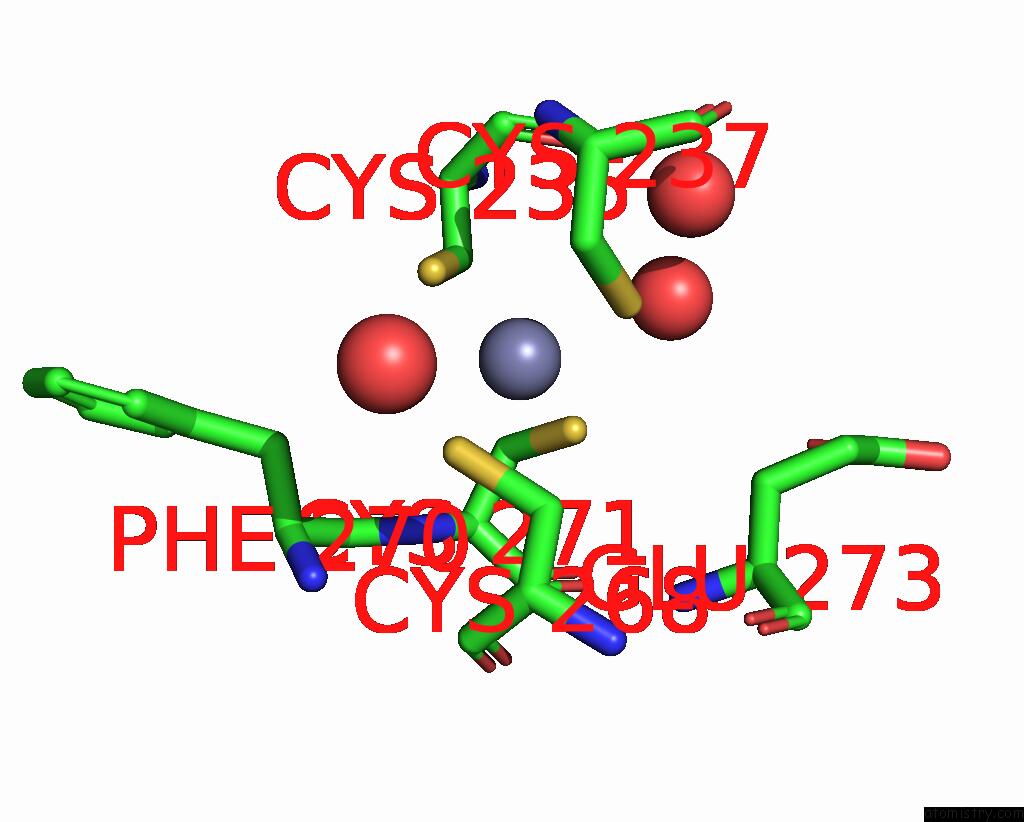
Mono view

Stereo pair view
Mono view
Stereo pair view
A full contact list of Zinc with other atoms in the Zn binding
site number 1 of Crystal Structure of the Bacillus Subtilis HSP33 within 5.0Å range:
|
Zinc binding site 2 out of 2 in 1vzy
Go back to
Zinc binding site 2 out
of 2 in the Crystal Structure of the Bacillus Subtilis HSP33

Mono view

Stereo pair view
Mono view
Stereo pair view
A full contact list of Zinc with other atoms in the Zn binding
site number 2 of Crystal Structure of the Bacillus Subtilis HSP33 within 5.0Å range:
|
Reference:
I.Janda,
Y.Devedjiev,
U.Derewenda,
Z.Dauter,
J.Bielnicki,
D.R.Cooper,
P.C.Graf,
A.Joachimiak,
U.Jakob,
Z.S.Derewenda.
The Crystal Structure of the Reduced, ZN2+-Bound Form of the B. Subtilis HSP33 Chaperone and Its Implications For the Activation Mechanism. Structure V. 12 1901 2004.
ISSN: ISSN 0969-2126
PubMed: 15458638
DOI: 10.1016/J.STR.2004.08.003
Page generated: Tue Aug 19 23:53:34 2025
ISSN: ISSN 0969-2126
PubMed: 15458638
DOI: 10.1016/J.STR.2004.08.003
Last articles
Fe in 9VR0Fe in 9UD8
Fe in 9QDT
Fe in 9S2T
Fe in 9JQA
Fe in 9IYV
Fe in 9J47
Fe in 9JQM
Fe in 9J2L
Fe in 9J1Q