Zinc »
PDB 1ul4-1v13 »
1urj »
Zinc in PDB 1urj: Single Stranded Dna-Binding Protein(ICP8) From Herpes Simplex Virus-1
Protein crystallography data
The structure of Single Stranded Dna-Binding Protein(ICP8) From Herpes Simplex Virus-1, PDB code: 1urj
was solved by
S.Panjikar,
M.Mapelli,
P.A.Tucker,
with X-Ray Crystallography technique. A brief refinement statistics is given in the table below:
| Resolution Low / High (Å) | 20.00 / 3.00 |
| Space group | P 21 21 21 |
| Cell size a, b, c (Å), α, β, γ (°) | 100.910, 145.370, 162.030, 90.00, 90.00, 90.00 |
| R / Rfree (%) | 23.5 / 28.6 |
Other elements in 1urj:
The structure of Single Stranded Dna-Binding Protein(ICP8) From Herpes Simplex Virus-1 also contains other interesting chemical elements:
| Mercury | (Hg) | 8 atoms |
Zinc Binding Sites:
The binding sites of Zinc atom in the Single Stranded Dna-Binding Protein(ICP8) From Herpes Simplex Virus-1
(pdb code 1urj). This binding sites where shown within
5.0 Angstroms radius around Zinc atom.
In total 2 binding sites of Zinc where determined in the Single Stranded Dna-Binding Protein(ICP8) From Herpes Simplex Virus-1, PDB code: 1urj:
Jump to Zinc binding site number: 1; 2;
In total 2 binding sites of Zinc where determined in the Single Stranded Dna-Binding Protein(ICP8) From Herpes Simplex Virus-1, PDB code: 1urj:
Jump to Zinc binding site number: 1; 2;
Zinc binding site 1 out of 2 in 1urj
Go back to
Zinc binding site 1 out
of 2 in the Single Stranded Dna-Binding Protein(ICP8) From Herpes Simplex Virus-1
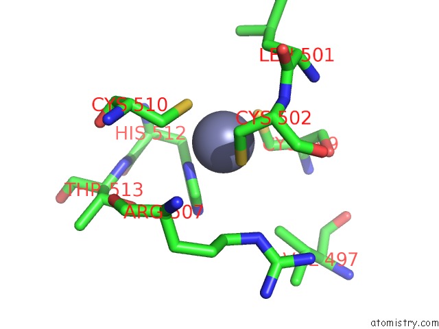
Mono view
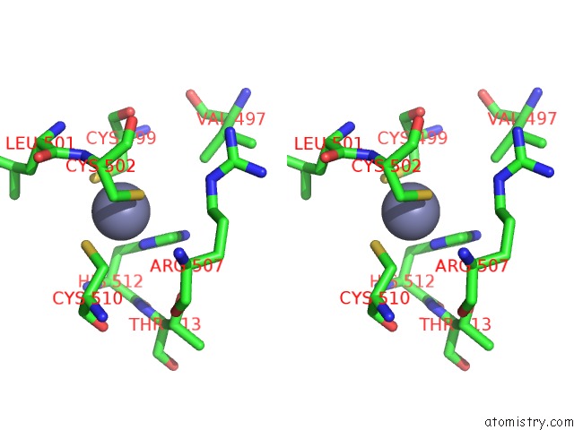
Stereo pair view
Mono view
Stereo pair view
A full contact list of Zinc with other atoms in the Zn binding
site number 1 of Single Stranded Dna-Binding Protein(ICP8) From Herpes Simplex Virus-1 within 5.0Å range:
|
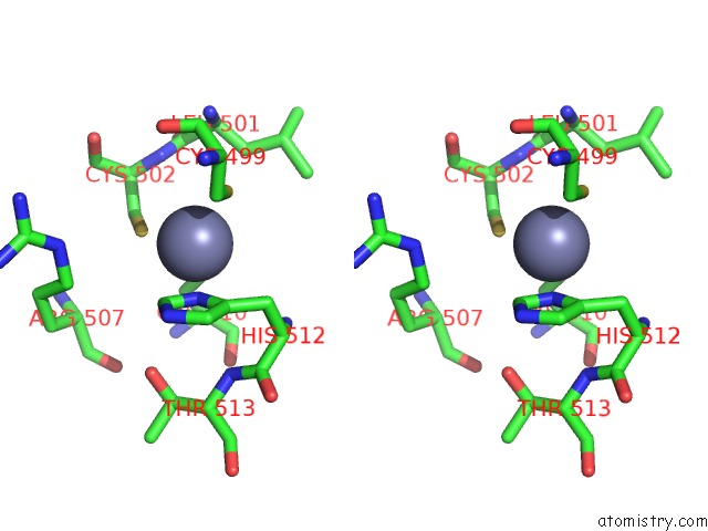
Zinc binding site 2 out of 2 in 1urj
Go back to
Zinc binding site 2 out
of 2 in the Single Stranded Dna-Binding Protein(ICP8) From Herpes Simplex Virus-1
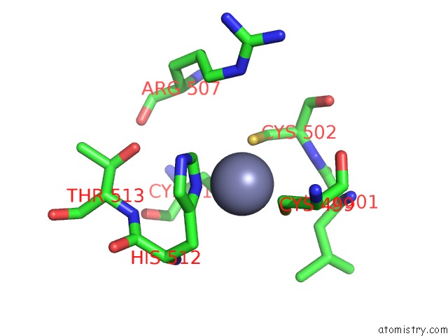
Mono view
Stereo pair view
Mono view
Stereo pair view
A full contact list of Zinc with other atoms in the Zn binding
site number 2 of Single Stranded Dna-Binding Protein(ICP8) From Herpes Simplex Virus-1 within 5.0Å range:
|
Reference:
M.Mapelli,
S.Panjikar,
P.A.Tucker.
The Crystal Structure of the Herpes Simplex Virus 1 Ssdna-Binding Protein Suggests the Structural Basis For Flexible, Cooperative Single-Stranded Dna Binding. J. Biol. Chem. V. 280 2990 2005.
ISSN: ISSN 0021-9258
PubMed: 15507432
DOI: 10.1074/JBC.M406780200
Page generated: Wed Oct 16 19:36:06 2024
ISSN: ISSN 0021-9258
PubMed: 15507432
DOI: 10.1074/JBC.M406780200
Last articles
K in 5THUK in 5T5I
K in 5THS
K in 5TF6
K in 5TD7
K in 5TCF
K in 5TAO
K in 5T5N
K in 5SDT
K in 5SCL