Zinc »
PDB 1ul4-1v13 »
1umt »
Zinc in PDB 1umt: Stromelysin-1 Catalytic Domain with Hydrophobic Inhibitor Bound, pH 7.0, 32OC, 20 Mm CACL2, 15% Acetonitrile; uc(Nmr) Average of 20 Structures Minimized with Restraints
Enzymatic activity of Stromelysin-1 Catalytic Domain with Hydrophobic Inhibitor Bound, pH 7.0, 32OC, 20 Mm CACL2, 15% Acetonitrile; uc(Nmr) Average of 20 Structures Minimized with Restraints
All present enzymatic activity of Stromelysin-1 Catalytic Domain with Hydrophobic Inhibitor Bound, pH 7.0, 32OC, 20 Mm CACL2, 15% Acetonitrile; uc(Nmr) Average of 20 Structures Minimized with Restraints:
3.4.24.17;
3.4.24.17;
Other elements in 1umt:
The structure of Stromelysin-1 Catalytic Domain with Hydrophobic Inhibitor Bound, pH 7.0, 32OC, 20 Mm CACL2, 15% Acetonitrile; uc(Nmr) Average of 20 Structures Minimized with Restraints also contains other interesting chemical elements:
| Calcium | (Ca) | 1 atom |
Zinc Binding Sites:
The binding sites of Zinc atom in the Stromelysin-1 Catalytic Domain with Hydrophobic Inhibitor Bound, pH 7.0, 32OC, 20 Mm CACL2, 15% Acetonitrile; uc(Nmr) Average of 20 Structures Minimized with Restraints
(pdb code 1umt). This binding sites where shown within
5.0 Angstroms radius around Zinc atom.
In total 2 binding sites of Zinc where determined in the Stromelysin-1 Catalytic Domain with Hydrophobic Inhibitor Bound, pH 7.0, 32OC, 20 Mm CACL2, 15% Acetonitrile; uc(Nmr) Average of 20 Structures Minimized with Restraints, PDB code: 1umt:
Jump to Zinc binding site number: 1; 2;
In total 2 binding sites of Zinc where determined in the Stromelysin-1 Catalytic Domain with Hydrophobic Inhibitor Bound, pH 7.0, 32OC, 20 Mm CACL2, 15% Acetonitrile; uc(Nmr) Average of 20 Structures Minimized with Restraints, PDB code: 1umt:
Jump to Zinc binding site number: 1; 2;
Zinc binding site 1 out of 2 in 1umt
Go back to
Zinc binding site 1 out
of 2 in the Stromelysin-1 Catalytic Domain with Hydrophobic Inhibitor Bound, pH 7.0, 32OC, 20 Mm CACL2, 15% Acetonitrile; uc(Nmr) Average of 20 Structures Minimized with Restraints
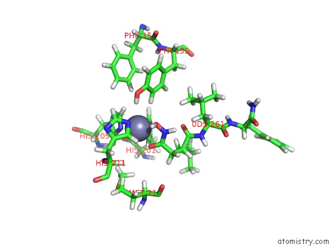
Mono view
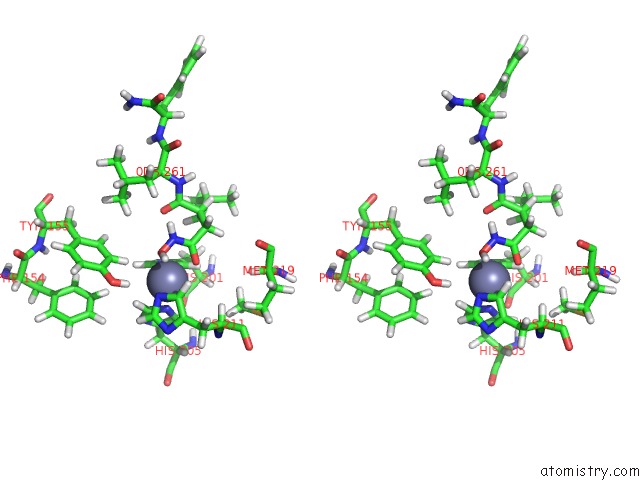
Stereo pair view
Mono view
Stereo pair view
A full contact list of Zinc with other atoms in the Zn binding
site number 1 of Stromelysin-1 Catalytic Domain with Hydrophobic Inhibitor Bound, pH 7.0, 32OC, 20 Mm CACL2, 15% Acetonitrile; uc(Nmr) Average of 20 Structures Minimized with Restraints within 5.0Å range:
|
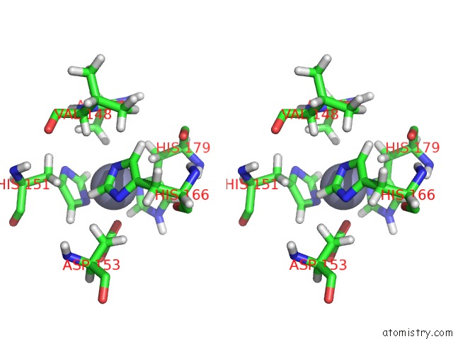
Zinc binding site 2 out of 2 in 1umt
Go back to
Zinc binding site 2 out
of 2 in the Stromelysin-1 Catalytic Domain with Hydrophobic Inhibitor Bound, pH 7.0, 32OC, 20 Mm CACL2, 15% Acetonitrile; uc(Nmr) Average of 20 Structures Minimized with Restraints
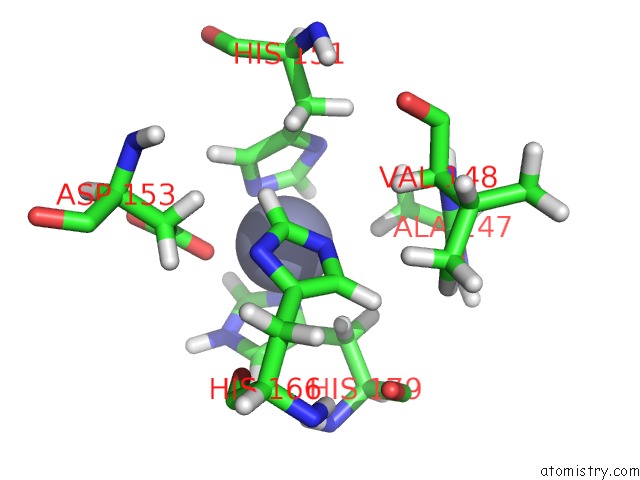
Mono view
Stereo pair view
Mono view
Stereo pair view
A full contact list of Zinc with other atoms in the Zn binding
site number 2 of Stromelysin-1 Catalytic Domain with Hydrophobic Inhibitor Bound, pH 7.0, 32OC, 20 Mm CACL2, 15% Acetonitrile; uc(Nmr) Average of 20 Structures Minimized with Restraints within 5.0Å range:
|
Reference:
S.R.Van Doren,
A.V.Kurochkin,
W.Hu,
Q.Z.Ye,
L.L.Johnson,
D.J.Hupe,
E.R.Zuiderweg.
Solution Structure of the Catalytic Domain of Human Stromelysin Complexed with A Hydrophobic Inhibitor. Protein Sci. V. 4 2487 1995.
ISSN: ISSN 0961-8368
PubMed: 8580839
Page generated: Wed Oct 16 19:33:52 2024
ISSN: ISSN 0961-8368
PubMed: 8580839
Last articles
K in 7QIXK in 7QNO
K in 7QIY
K in 7Q3X
K in 7QDN
K in 7QF6
K in 7Q0G
K in 7Q1C
K in 7Q1B
K in 7PXH