Zinc »
PDB 1u2n-1uip »
1ue1 »
Zinc in PDB 1ue1: Crystal Structure of the Single-Stranded Dna-Binding Protein From Mycobacterium Tuberculosis
Protein crystallography data
The structure of Crystal Structure of the Single-Stranded Dna-Binding Protein From Mycobacterium Tuberculosis, PDB code: 1ue1
was solved by
K.Saikrishnan,
J.Jeyakanthan,
J.Venkatesh,
N.Acharya,
K.Sekar,
U.Varshney,
M.Vijayan,
Tb Structural Genomics Consortium(Tbsgc),
with X-Ray Crystallography technique. A brief refinement statistics is given in the table below:
| Resolution Low / High (Å) | 15.00 / 2.50 |
| Space group | P 31 2 1 |
| Cell size a, b, c (Å), α, β, γ (°) | 78.720, 78.720, 77.160, 90.00, 90.00, 120.00 |
| R / Rfree (%) | 22.8 / 28.8 |
Zinc Binding Sites:
The binding sites of Zinc atom in the Crystal Structure of the Single-Stranded Dna-Binding Protein From Mycobacterium Tuberculosis
(pdb code 1ue1). This binding sites where shown within
5.0 Angstroms radius around Zinc atom.
In total only one binding site of Zinc was determined in the Crystal Structure of the Single-Stranded Dna-Binding Protein From Mycobacterium Tuberculosis, PDB code: 1ue1:
In total only one binding site of Zinc was determined in the Crystal Structure of the Single-Stranded Dna-Binding Protein From Mycobacterium Tuberculosis, PDB code: 1ue1:
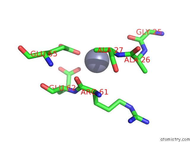
Zinc binding site 1 out of 1 in 1ue1
Go back to
Zinc binding site 1 out
of 1 in the Crystal Structure of the Single-Stranded Dna-Binding Protein From Mycobacterium Tuberculosis
Mono view

Stereo pair view
Mono view
Stereo pair view
A full contact list of Zinc with other atoms in the Zn binding
site number 1 of Crystal Structure of the Single-Stranded Dna-Binding Protein From Mycobacterium Tuberculosis within 5.0Å range:
|
Reference:
K.Saikrishnan,
J.Jeyakanthan,
J.Venkatesh,
N.Acharya,
K.Sekar,
U.Varshney,
M.Vijayan.
Structure of Mycobacterium Tuberculosis Single-Stranded Dna-Binding Protein. Variability in Quaternary Structure and Its Implications J.Mol.Biol. V. 331 385 2003.
ISSN: ISSN 0022-2836
PubMed: 12888346
DOI: 10.1016/S0022-2836(03)00729-0
Page generated: Wed Oct 16 19:30:28 2024
ISSN: ISSN 0022-2836
PubMed: 12888346
DOI: 10.1016/S0022-2836(03)00729-0
Last articles
Mg in 5XTMMg in 5XUT
Mg in 5XUS
Mg in 5XUJ
Mg in 5XUI
Mg in 5XU1
Mg in 5XT8
Mg in 5XT2
Mg in 5XR7
Mg in 5XR6