Zinc »
PDB 1u2n-1uip »
1u3v »
Zinc in PDB 1u3v: Crystal Structure of Human Alcohol Dehydrogenase Beta-1-Beta-1 Isoform Complexed with N-Heptylformamide Determined to 1.65 Angstrom Resolution
Enzymatic activity of Crystal Structure of Human Alcohol Dehydrogenase Beta-1-Beta-1 Isoform Complexed with N-Heptylformamide Determined to 1.65 Angstrom Resolution
All present enzymatic activity of Crystal Structure of Human Alcohol Dehydrogenase Beta-1-Beta-1 Isoform Complexed with N-Heptylformamide Determined to 1.65 Angstrom Resolution:
1.1.1.1;
1.1.1.1;
Protein crystallography data
The structure of Crystal Structure of Human Alcohol Dehydrogenase Beta-1-Beta-1 Isoform Complexed with N-Heptylformamide Determined to 1.65 Angstrom Resolution, PDB code: 1u3v
was solved by
B.J.Gibbons,
T.D.Hurley,
with X-Ray Crystallography technique. A brief refinement statistics is given in the table below:
| Resolution Low / High (Å) | 30.00 / 1.65 |
| Space group | P 1 |
| Cell size a, b, c (Å), α, β, γ (°) | 44.320, 52.830, 90.350, 79.58, 89.43, 68.60 |
| R / Rfree (%) | 16.4 / 18.5 |
Zinc Binding Sites:
The binding sites of Zinc atom in the Crystal Structure of Human Alcohol Dehydrogenase Beta-1-Beta-1 Isoform Complexed with N-Heptylformamide Determined to 1.65 Angstrom Resolution
(pdb code 1u3v). This binding sites where shown within
5.0 Angstroms radius around Zinc atom.
In total 4 binding sites of Zinc where determined in the Crystal Structure of Human Alcohol Dehydrogenase Beta-1-Beta-1 Isoform Complexed with N-Heptylformamide Determined to 1.65 Angstrom Resolution, PDB code: 1u3v:
Jump to Zinc binding site number: 1; 2; 3; 4;
In total 4 binding sites of Zinc where determined in the Crystal Structure of Human Alcohol Dehydrogenase Beta-1-Beta-1 Isoform Complexed with N-Heptylformamide Determined to 1.65 Angstrom Resolution, PDB code: 1u3v:
Jump to Zinc binding site number: 1; 2; 3; 4;
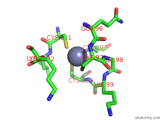
Zinc binding site 1 out of 4 in 1u3v
Go back to
Zinc binding site 1 out
of 4 in the Crystal Structure of Human Alcohol Dehydrogenase Beta-1-Beta-1 Isoform Complexed with N-Heptylformamide Determined to 1.65 Angstrom Resolution
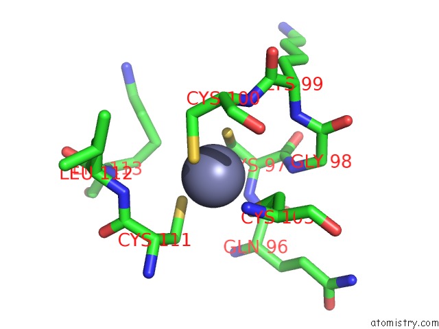
Mono view
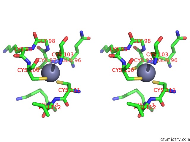
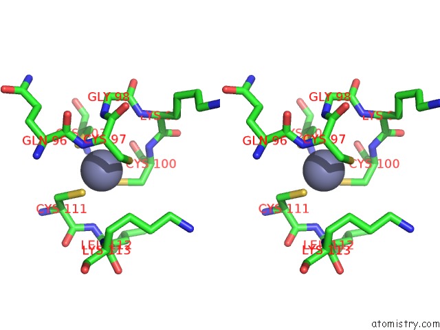
Stereo pair view
Mono view
Stereo pair view
A full contact list of Zinc with other atoms in the Zn binding
site number 1 of Crystal Structure of Human Alcohol Dehydrogenase Beta-1-Beta-1 Isoform Complexed with N-Heptylformamide Determined to 1.65 Angstrom Resolution within 5.0Å range:
|
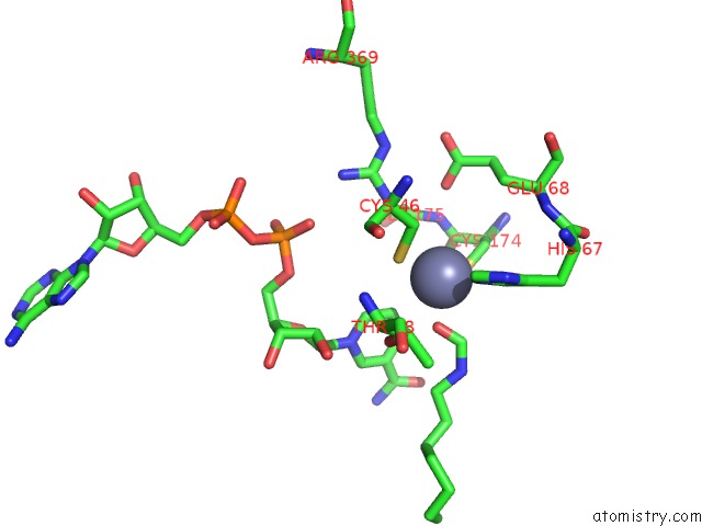
Zinc binding site 2 out of 4 in 1u3v
Go back to
Zinc binding site 2 out
of 4 in the Crystal Structure of Human Alcohol Dehydrogenase Beta-1-Beta-1 Isoform Complexed with N-Heptylformamide Determined to 1.65 Angstrom Resolution
Mono view
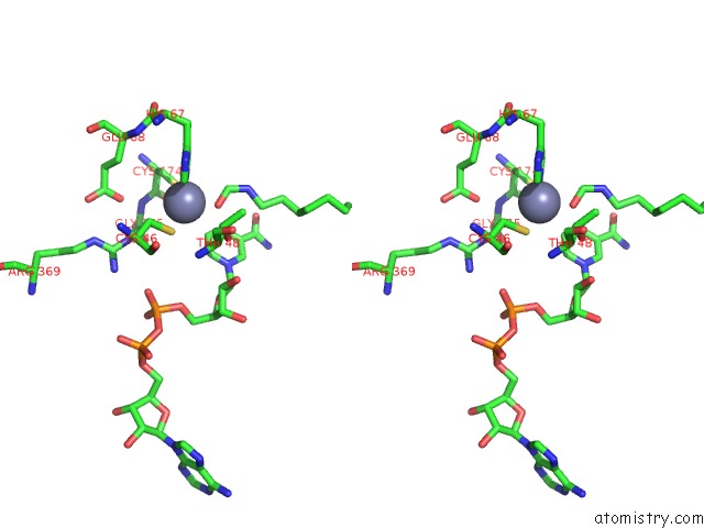
Stereo pair view
Mono view
Stereo pair view
A full contact list of Zinc with other atoms in the Zn binding
site number 2 of Crystal Structure of Human Alcohol Dehydrogenase Beta-1-Beta-1 Isoform Complexed with N-Heptylformamide Determined to 1.65 Angstrom Resolution within 5.0Å range:
|
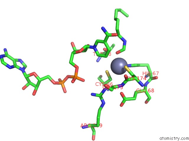
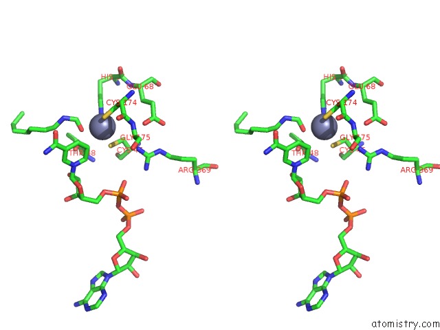
Zinc binding site 3 out of 4 in 1u3v
Go back to
Zinc binding site 3 out
of 4 in the Crystal Structure of Human Alcohol Dehydrogenase Beta-1-Beta-1 Isoform Complexed with N-Heptylformamide Determined to 1.65 Angstrom Resolution
Mono view
Stereo pair view
Mono view
Stereo pair view
A full contact list of Zinc with other atoms in the Zn binding
site number 3 of Crystal Structure of Human Alcohol Dehydrogenase Beta-1-Beta-1 Isoform Complexed with N-Heptylformamide Determined to 1.65 Angstrom Resolution within 5.0Å range:
|
Zinc binding site 4 out of 4 in 1u3v
Go back to
Zinc binding site 4 out
of 4 in the Crystal Structure of Human Alcohol Dehydrogenase Beta-1-Beta-1 Isoform Complexed with N-Heptylformamide Determined to 1.65 Angstrom Resolution
Mono view
Stereo pair view
Mono view
Stereo pair view
A full contact list of Zinc with other atoms in the Zn binding
site number 4 of Crystal Structure of Human Alcohol Dehydrogenase Beta-1-Beta-1 Isoform Complexed with N-Heptylformamide Determined to 1.65 Angstrom Resolution within 5.0Å range:
|
Reference:
B.J.Gibbons,
T.D.Hurley.
Structure of Three Class I Human Alcohol Dehydrogenases Complexed with Isoenzyme Specific Formamide Inhibitors Biochemistry V. 43 12555 2004.
ISSN: ISSN 0006-2960
PubMed: 15449945
DOI: 10.1021/BI0489107
Page generated: Wed Oct 16 19:25:59 2024
ISSN: ISSN 0006-2960
PubMed: 15449945
DOI: 10.1021/BI0489107
Last articles
Mg in 5SEYMg in 5SEX
Mg in 5SEV
Mg in 5SEU
Mg in 5SEW
Mg in 5SET
Mg in 5SES
Mg in 5SER
Mg in 5SEQ
Mg in 5SEO