Zinc »
PDB 1to5-1u22 »
1u05 »
Zinc in PDB 1u05: Crystal Structure of Protein Yfih From Shigella Flexneri, Pfam DUF152
Protein crystallography data
The structure of Crystal Structure of Protein Yfih From Shigella Flexneri, Pfam DUF152, PDB code: 1u05
was solved by
J.Seetharaman,
S.Swaminathan,
S.K.Burley,
New York Sgx Research Centerfor Structural Genomics (Nysgxrc),
with X-Ray Crystallography technique. A brief refinement statistics is given in the table below:
| Resolution Low / High (Å) | 33.00 / 2.50 |
| Space group | P 1 |
| Cell size a, b, c (Å), α, β, γ (°) | 43.683, 50.465, 55.265, 90.25, 96.51, 90.29 |
| R / Rfree (%) | 21.7 / 28.3 |
Zinc Binding Sites:
The binding sites of Zinc atom in the Crystal Structure of Protein Yfih From Shigella Flexneri, Pfam DUF152
(pdb code 1u05). This binding sites where shown within
5.0 Angstroms radius around Zinc atom.
In total 2 binding sites of Zinc where determined in the Crystal Structure of Protein Yfih From Shigella Flexneri, Pfam DUF152, PDB code: 1u05:
Jump to Zinc binding site number: 1; 2;
In total 2 binding sites of Zinc where determined in the Crystal Structure of Protein Yfih From Shigella Flexneri, Pfam DUF152, PDB code: 1u05:
Jump to Zinc binding site number: 1; 2;
Zinc binding site 1 out of 2 in 1u05
Go back to
Zinc binding site 1 out
of 2 in the Crystal Structure of Protein Yfih From Shigella Flexneri, Pfam DUF152
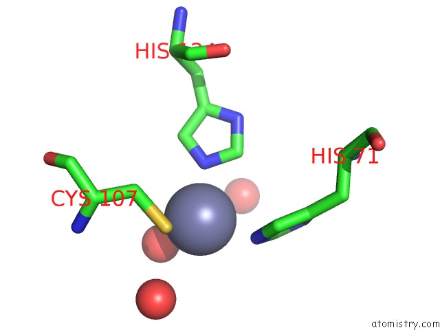
Mono view
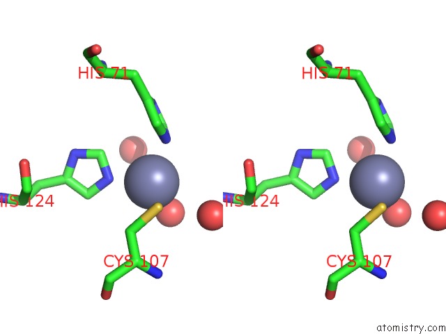
Stereo pair view
Mono view
Stereo pair view
A full contact list of Zinc with other atoms in the Zn binding
site number 1 of Crystal Structure of Protein Yfih From Shigella Flexneri, Pfam DUF152 within 5.0Å range:
|
Zinc binding site 2 out of 2 in 1u05
Go back to
Zinc binding site 2 out
of 2 in the Crystal Structure of Protein Yfih From Shigella Flexneri, Pfam DUF152
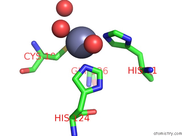
Mono view
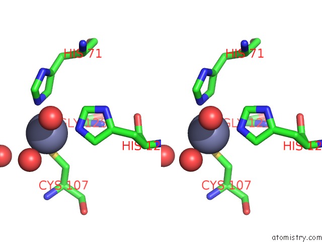
Stereo pair view
Mono view
Stereo pair view
A full contact list of Zinc with other atoms in the Zn binding
site number 2 of Crystal Structure of Protein Yfih From Shigella Flexneri, Pfam DUF152 within 5.0Å range:
|
Reference:
J.Seetharaman,
S.Swaminathan.
Crystal Structure of Conserved Hypothetical Protein To Be Published.
Page generated: Wed Oct 16 19:22:26 2024
Last articles
Mg in 2BIFMg in 2BIA
Mg in 2BI5
Mg in 2BI9
Mg in 2BI3
Mg in 2BI2
Mg in 2BI1
Mg in 2BHZ
Mg in 2BHX
Mg in 2BHD