Zinc »
PDB 1sdz-1sxb »
1slm »
Zinc in PDB 1slm: Crystal Structure of Fibroblast Stromelysin-1: the C-Truncated Human Proenzyme
Enzymatic activity of Crystal Structure of Fibroblast Stromelysin-1: the C-Truncated Human Proenzyme
All present enzymatic activity of Crystal Structure of Fibroblast Stromelysin-1: the C-Truncated Human Proenzyme:
3.4.24.17;
3.4.24.17;
Protein crystallography data
The structure of Crystal Structure of Fibroblast Stromelysin-1: the C-Truncated Human Proenzyme, PDB code: 1slm
was solved by
J.W.Becker,
with X-Ray Crystallography technique. A brief refinement statistics is given in the table below:
| Resolution Low / High (Å) | 20.00 / 1.90 |
| Space group | F 2 2 2 |
| Cell size a, b, c (Å), α, β, γ (°) | 111.040, 145.560, 76.750, 90.00, 90.00, 90.00 |
| R / Rfree (%) | 21.9 / 25.6 |
Other elements in 1slm:
The structure of Crystal Structure of Fibroblast Stromelysin-1: the C-Truncated Human Proenzyme also contains other interesting chemical elements:
| Calcium | (Ca) | 2 atoms |
Zinc Binding Sites:
The binding sites of Zinc atom in the Crystal Structure of Fibroblast Stromelysin-1: the C-Truncated Human Proenzyme
(pdb code 1slm). This binding sites where shown within
5.0 Angstroms radius around Zinc atom.
In total 2 binding sites of Zinc where determined in the Crystal Structure of Fibroblast Stromelysin-1: the C-Truncated Human Proenzyme, PDB code: 1slm:
Jump to Zinc binding site number: 1; 2;
In total 2 binding sites of Zinc where determined in the Crystal Structure of Fibroblast Stromelysin-1: the C-Truncated Human Proenzyme, PDB code: 1slm:
Jump to Zinc binding site number: 1; 2;
Zinc binding site 1 out of 2 in 1slm
Go back to
Zinc binding site 1 out
of 2 in the Crystal Structure of Fibroblast Stromelysin-1: the C-Truncated Human Proenzyme
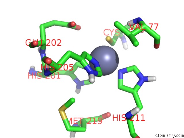
Mono view
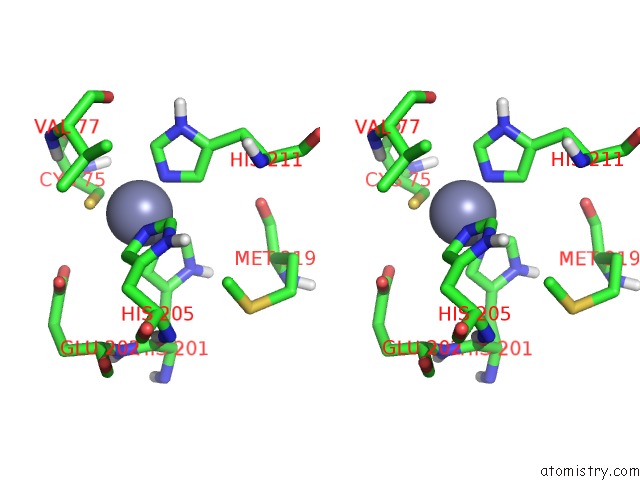
Stereo pair view
Mono view
Stereo pair view
A full contact list of Zinc with other atoms in the Zn binding
site number 1 of Crystal Structure of Fibroblast Stromelysin-1: the C-Truncated Human Proenzyme within 5.0Å range:
|
Zinc binding site 2 out of 2 in 1slm
Go back to
Zinc binding site 2 out
of 2 in the Crystal Structure of Fibroblast Stromelysin-1: the C-Truncated Human Proenzyme
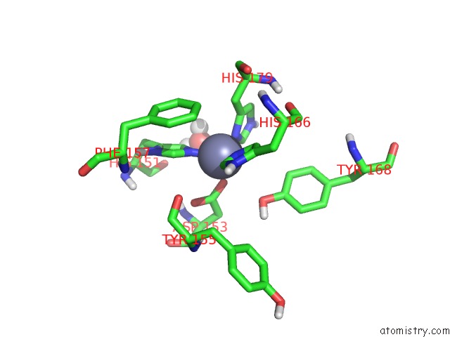
Mono view
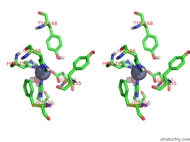
Stereo pair view
Mono view
Stereo pair view
A full contact list of Zinc with other atoms in the Zn binding
site number 2 of Crystal Structure of Fibroblast Stromelysin-1: the C-Truncated Human Proenzyme within 5.0Å range:
|
Reference:
J.W.Becker,
A.I.Marcy,
L.L.Rokosz,
M.G.Axel,
J.J.Burbaum,
P.M.Fitzgerald,
P.M.Cameron,
C.K.Esser,
W.K.Hagmann,
J.D.Hermes,
J.P.Springer.
Stromelysin-1: Three-Dimensional Structure of the Inhibited Catalytic Domain and of the C-Truncated Proenzyme. Protein Sci. V. 4 1966 1995.
ISSN: ISSN 0961-8368
PubMed: 8535233
Page generated: Wed Oct 16 18:52:06 2024
ISSN: ISSN 0961-8368
PubMed: 8535233
Last articles
Na in 7EAFNa in 7E50
Na in 7E5J
Na in 7E4C
Na in 7E21
Na in 7E1Z
Na in 7E4A
Na in 7E02
Na in 7DTF
Na in 7DTB