Atomistry » Zinc » PDB 1r2y-1raf » 1r3n
Atomistry »
  Zinc »
    PDB 1r2y-1raf »
      1r3n »

Zinc in PDB 1r3n: Crystal Structure of Beta-Alanine Synthase From Saccharomyces Kluyveri

Enzymatic activity of Crystal Structure of Beta-Alanine Synthase From Saccharomyces Kluyveri

All present enzymatic activity of Crystal Structure of Beta-Alanine Synthase From Saccharomyces Kluyveri:
3.5.1.6;

Protein crystallography data

The structure of Crystal Structure of Beta-Alanine Synthase From Saccharomyces Kluyveri, PDB code: 1r3n was solved by S.Lundgren, Z.Gojkovic, J.Piskur, D.Dobritzsch, with X-Ray Crystallography technique. A brief refinement statistics is given in the table below:

Resolution Low / High (Å) 25.00 / 2.70
Space group P 1 21 1
Cell size a, b, c (Å), α, β, γ (°) 117.230, 77.120, 225.520, 90.00, 95.05, 90.00
R / Rfree (%) 20.8 / 26.6

Zinc Binding Sites:

Pages:

>>> Page 1 <<< Page 2, Binding sites: 11 - 16;

Binding sites:

The binding sites of Zinc atom in the Crystal Structure of Beta-Alanine Synthase From Saccharomyces Kluyveri (pdb code 1r3n). This binding sites where shown within 5.0 Angstroms radius around Zinc atom.
In total 16 binding sites of Zinc where determined in the Crystal Structure of Beta-Alanine Synthase From Saccharomyces Kluyveri, PDB code: 1r3n:
Jump to Zinc binding site number: 1; 2; 3; 4; 5; 6; 7; 8; 9; 10;

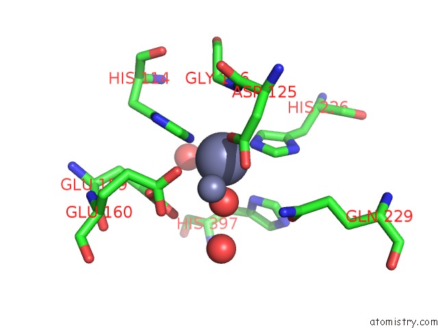
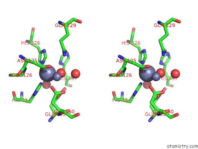
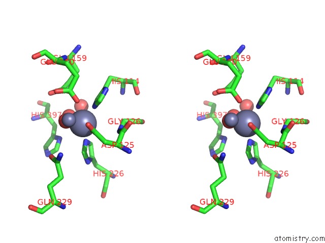
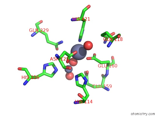
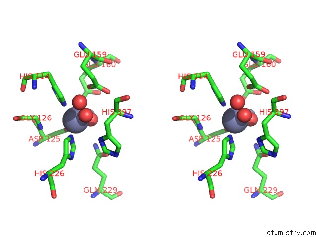
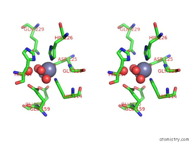
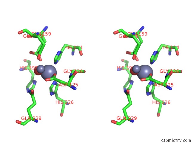
Zinc binding site 1 out of 16 in 1r3n

Go back to Zinc Binding Sites List in 1r3n
Zinc binding site 1 out of 16 in the Crystal Structure of Beta-Alanine Synthase From Saccharomyces Kluyveri


Mono view


Stereo pair view

A full contact list of Zinc with other atoms in the Zn binding site number 1 of Crystal Structure of Beta-Alanine Synthase From Saccharomyces Kluyveri within 5.0Å range:
probe atom residue distance (Å) B Occ
A:Zn500

b:15.5
occ:1.00
OD1 A:ASP125 2.0 18.2 1.0
NE2 A:HIS114 2.1 15.5 1.0
NE2 A:HIS226 2.1 14.4 1.0
O A:HOH2504 2.1 26.8 1.0
O A:HOH2503 2.3 17.8 1.0
CE1 A:HIS114 2.7 15.8 1.0
CE1 A:HIS226 2.8 13.4 1.0
CG A:ASP125 3.1 17.0 1.0
CD2 A:HIS226 3.3 14.9 1.0
CD2 A:HIS114 3.3 16.0 1.0
ZN A:ZN501 3.3 18.6 1.0
OD2 A:ASP125 3.3 19.4 1.0
N A:GLY126 3.8 15.8 1.0
ND1 A:HIS114 4.0 14.5 1.0
ND1 A:HIS226 4.0 13.0 1.0
OE2 A:GLU159 4.1 25.2 1.0
OE1 A:GLU159 4.1 25.4 1.0
CD A:GLU159 4.2 23.4 1.0
CA A:GLY126 4.2 15.9 1.0
CG A:HIS226 4.3 14.4 1.0
CG A:HIS114 4.3 15.5 1.0
ND1 A:HIS397 4.3 13.5 1.0
OE2 A:GLU160 4.4 18.2 1.0
C A:ASP125 4.4 15.9 1.0
CB A:ASP125 4.4 16.2 1.0
O A:HOH2505 4.4 24.8 1.0
O A:HOH2508 4.6 42.3 1.0
CE1 A:HIS397 4.6 13.6 1.0
OE1 A:GLN229 4.7 19.3 1.0
CA A:ASP125 4.7 16.0 1.0
CD A:GLU160 4.8 18.8 1.0
OE1 A:GLU160 4.9 18.9 1.0
NE2 A:GLN229 4.9 16.9 1.0
CD A:GLN229 5.0 16.6 1.0

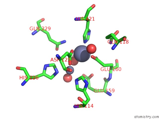
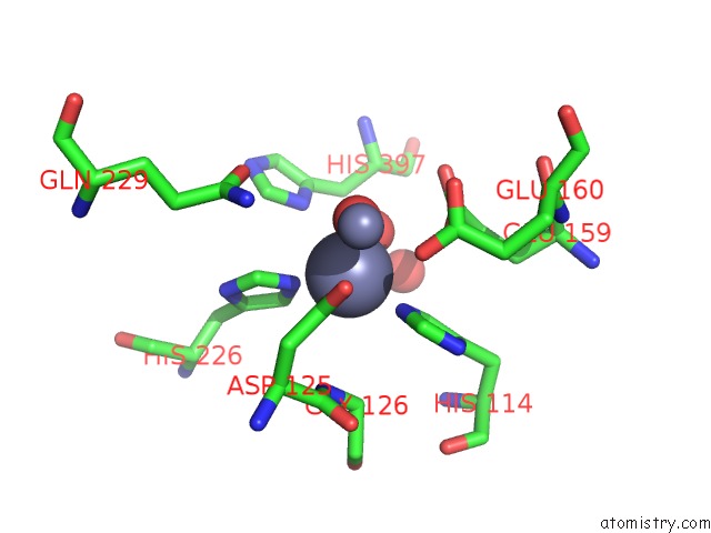
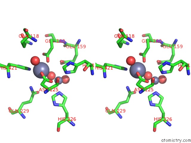
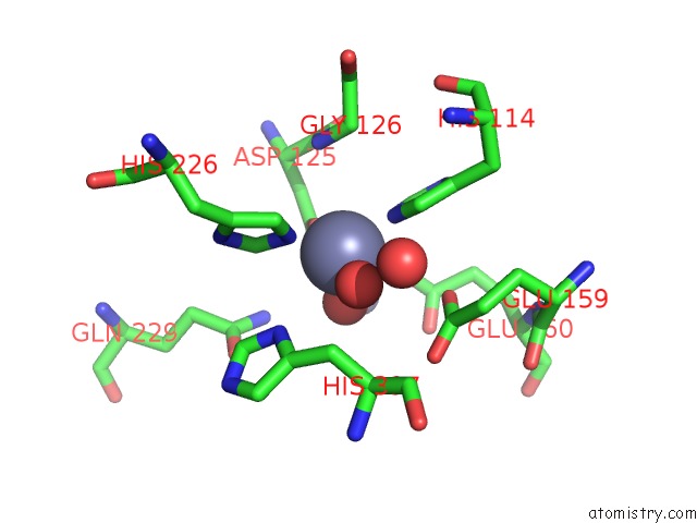
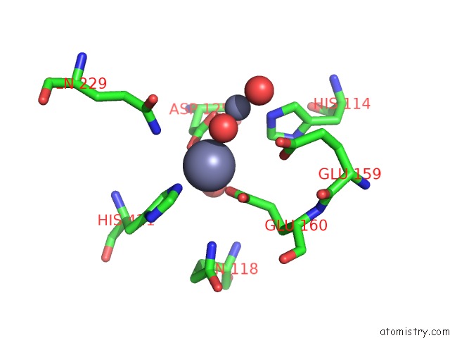
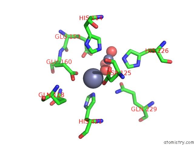
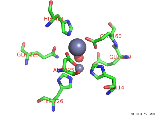
Zinc binding site 2 out of 16 in 1r3n

Go back to Zinc Binding Sites List in 1r3n
Zinc binding site 2 out of 16 in the Crystal Structure of Beta-Alanine Synthase From Saccharomyces Kluyveri


Mono view


Stereo pair view

A full contact list of Zinc with other atoms in the Zn binding site number 2 of Crystal Structure of Beta-Alanine Synthase From Saccharomyces Kluyveri within 5.0Å range:
probe atom residue distance (Å) B Occ
A:Zn501

b:18.6
occ:1.00
OD2 A:ASP125 2.0 19.4 1.0
OE2 A:GLU160 2.0 18.2 1.0
NE2 A:HIS421 2.1 18.8 1.0
O A:HOH2503 2.5 17.8 1.0
CD A:GLU160 2.6 18.8 1.0
OE1 A:GLU160 2.7 18.9 1.0
CG A:ASP125 2.8 17.0 1.0
CD2 A:HIS421 2.9 17.8 1.0
OD1 A:ASP125 3.0 18.2 1.0
CE1 A:HIS421 3.1 19.9 1.0
ZN A:ZN500 3.3 15.5 1.0
NE2 A:GLN229 3.6 16.9 1.0
O A:HOH2506 3.8 12.5 1.0
CE1 A:HIS114 4.0 15.8 1.0
CG A:GLU160 4.1 16.8 1.0
CB A:ASP125 4.1 16.2 1.0
CG A:HIS421 4.1 17.7 1.0
O A:HOH2508 4.2 42.3 1.0
ND1 A:HIS421 4.2 19.3 1.0
OE1 A:GLU159 4.3 25.4 1.0
NE2 A:HIS114 4.4 15.5 1.0
CD A:GLN229 4.6 16.6 1.0
OE1 A:GLN229 4.8 19.3 1.0
CG A:GLN118 4.8 15.7 1.0
OE2 A:GLU159 4.9 25.2 1.0
CE1 A:HIS226 4.9 13.4 1.0
O A:HOH2504 5.0 26.8 1.0
CD A:GLU159 5.0 23.4 1.0

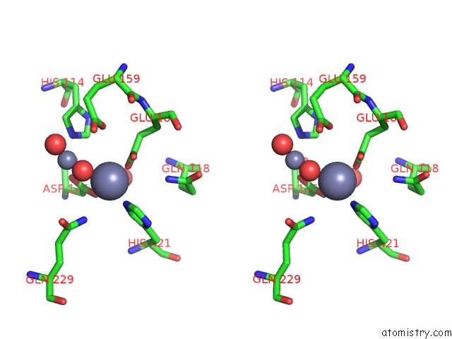
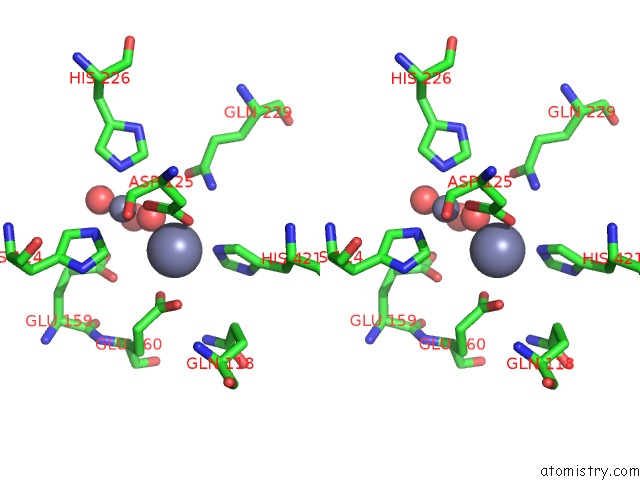
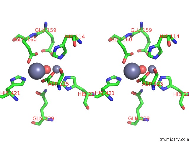
Zinc binding site 3 out of 16 in 1r3n

Go back to Zinc Binding Sites List in 1r3n
Zinc binding site 3 out of 16 in the Crystal Structure of Beta-Alanine Synthase From Saccharomyces Kluyveri


Mono view


Stereo pair view

A full contact list of Zinc with other atoms in the Zn binding site number 3 of Crystal Structure of Beta-Alanine Synthase From Saccharomyces Kluyveri within 5.0Å range:
probe atom residue distance (Å) B Occ
B:Zn500

b:16.2
occ:1.00
O B:HOH1506 1.9 18.1 1.0
OD2 B:ASP125 2.0 16.3 1.0
NE2 B:HIS114 2.1 15.6 1.0
NE2 B:HIS226 2.1 15.1 1.0
O B:HOH1503 2.4 11.5 1.0
CE1 B:HIS226 2.9 14.8 1.0
CE1 B:HIS114 3.0 14.8 1.0
CD2 B:HIS114 3.1 15.2 1.0
CG B:ASP125 3.2 16.5 1.0
CD2 B:HIS226 3.2 15.1 1.0
ZN B:ZN501 3.4 18.7 1.0
OE1 B:GLU159 3.5 22.2 1.0
OD1 B:ASP125 3.7 18.7 1.0
N B:GLY126 3.8 15.8 1.0
OE2 B:GLU160 3.8 18.3 1.0
ND1 B:HIS397 4.0 14.1 1.0
CD B:GLU159 4.1 21.6 1.0
ND1 B:HIS226 4.1 15.0 1.0
ND1 B:HIS114 4.1 15.8 1.0
CA B:GLY126 4.2 15.8 1.0
CG B:HIS114 4.2 15.4 1.0
CG B:HIS226 4.3 15.3 1.0
C B:ASP125 4.4 15.9 1.0
CB B:ASP125 4.5 16.0 1.0
CE1 B:HIS397 4.5 14.7 1.0
O B:HOH1504 4.5 24.5 1.0
OE2 B:GLU159 4.6 23.3 1.0
OE1 B:GLN229 4.7 19.5 1.0
CA B:ASP125 4.7 15.9 1.0
CD B:GLU160 4.7 16.2 1.0
CG B:GLU159 4.8 18.2 1.0
NE2 B:GLN229 4.9 18.5 1.0
CD B:GLN229 5.0 18.5 1.0

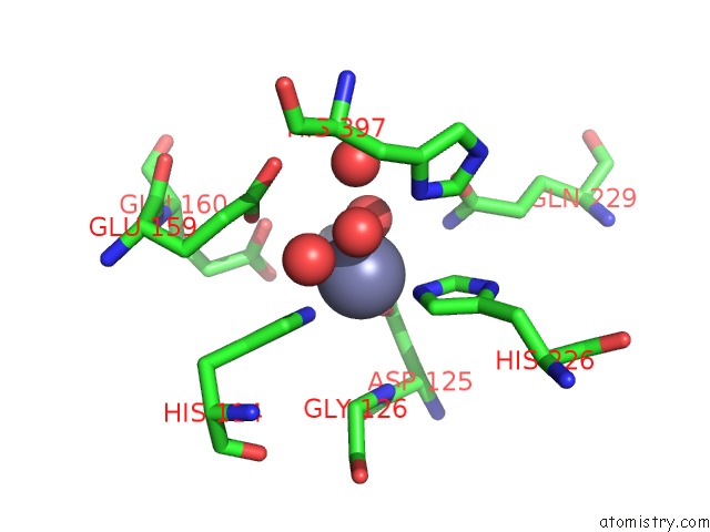
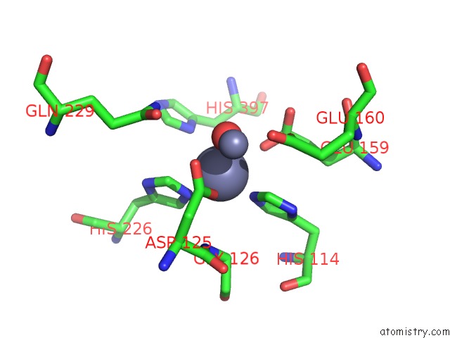
Zinc binding site 4 out of 16 in 1r3n

Go back to Zinc Binding Sites List in 1r3n
Zinc binding site 4 out of 16 in the Crystal Structure of Beta-Alanine Synthase From Saccharomyces Kluyveri


Mono view


Stereo pair view

A full contact list of Zinc with other atoms in the Zn binding site number 4 of Crystal Structure of Beta-Alanine Synthase From Saccharomyces Kluyveri within 5.0Å range:
probe atom residue distance (Å) B Occ
B:Zn501

b:18.7
occ:1.00
OD1 B:ASP125 2.0 18.7 1.0
OE2 B:GLU160 2.0 18.3 1.0
NE2 B:HIS421 2.1 19.0 1.0
O B:HOH1506 2.3 18.1 1.0
CG B:ASP125 2.8 16.5 1.0
CD B:GLU160 2.8 16.2 1.0
CD2 B:HIS421 2.9 18.0 1.0
OD2 B:ASP125 2.9 16.3 1.0
OE1 B:GLU160 3.0 15.9 1.0
CE1 B:HIS421 3.2 19.8 1.0
ZN B:ZN500 3.4 16.2 1.0
NE2 B:GLN229 3.5 18.5 1.0
O B:HOH1505 3.7 24.9 1.0
OE1 B:GLU159 4.0 22.2 1.0
CB B:ASP125 4.1 16.0 1.0
CG B:HIS421 4.1 17.5 1.0
CE1 B:HIS114 4.2 14.8 1.0
ND1 B:HIS421 4.2 19.4 1.0
CG B:GLU160 4.3 16.4 1.0
NE2 B:HIS114 4.3 15.6 1.0
CD B:GLN229 4.4 18.5 1.0
OE1 B:GLN229 4.5 19.5 1.0
NE2 B:HIS226 4.9 15.1 1.0
CE1 B:HIS226 4.9 14.8 1.0
O B:HOH1503 4.9 11.5 1.0
CD B:GLU159 4.9 21.6 1.0
CG B:GLN118 5.0 17.2 1.0
NE2 B:GLN118 5.0 18.1 1.0

Zinc binding site 5 out of 16 in 1r3n

Go back to Zinc Binding Sites List in 1r3n
Zinc binding site 5 out of 16 in the Crystal Structure of Beta-Alanine Synthase From Saccharomyces Kluyveri


Mono view


Stereo pair view

A full contact list of Zinc with other atoms in the Zn binding site number 5 of Crystal Structure of Beta-Alanine Synthase From Saccharomyces Kluyveri within 5.0Å range:
probe atom residue distance (Å) B Occ
C:Zn500

b:16.1
occ:1.00
O C:HOH3503 1.8 17.8 1.0
OD2 C:ASP125 2.0 16.4 1.0
NE2 C:HIS114 2.1 11.8 1.0
NE2 C:HIS226 2.1 11.5 1.0
O C:HOH3504 2.1 14.6 1.0
CE1 C:HIS226 2.8 10.9 1.0
CE1 C:HIS114 2.9 11.8 1.0
CG C:ASP125 3.1 16.4 1.0
CD2 C:HIS114 3.2 12.7 1.0
CD2 C:HIS226 3.3 11.2 1.0
ZN C:ZN501 3.4 18.4 1.0
OD1 C:ASP125 3.5 18.5 1.0
OE1 C:GLU159 3.7 27.1 1.0
N C:GLY126 3.7 15.8 1.0
ND1 C:HIS226 4.1 11.0 1.0
ND1 C:HIS114 4.1 12.2 1.0
CA C:GLY126 4.1 15.9 1.0
CD C:GLU159 4.2 25.6 1.0
CG C:HIS114 4.2 13.3 1.0
CG C:HIS226 4.3 12.2 1.0
O C:HOH3505 4.3 28.8 1.0
C C:ASP125 4.3 15.8 1.0
ND1 C:HIS397 4.4 14.6 1.0
CB C:ASP125 4.4 16.0 1.0
OE2 C:GLU159 4.5 27.3 1.0
NE2 C:GLN229 4.5 13.4 1.0
OE2 C:GLU160 4.5 18.6 1.0
CA C:ASP125 4.7 16.0 1.0
CD C:GLU160 4.8 18.6 1.0
OE1 C:GLU160 4.9 19.7 1.0
CD C:GLN229 4.9 13.3 1.0
CE1 C:HIS397 4.9 15.6 1.0
OE1 C:GLN229 5.0 10.7 1.0

Zinc binding site 6 out of 16 in 1r3n

Go back to Zinc Binding Sites List in 1r3n
Zinc binding site 6 out of 16 in the Crystal Structure of Beta-Alanine Synthase From Saccharomyces Kluyveri


Mono view


Stereo pair view

A full contact list of Zinc with other atoms in the Zn binding site number 6 of Crystal Structure of Beta-Alanine Synthase From Saccharomyces Kluyveri within 5.0Å range:
probe atom residue distance (Å) B Occ
C:Zn501

b:18.4
occ:1.00
OD1 C:ASP125 2.0 18.5 1.0
OE2 C:GLU160 2.0 18.6 1.0
NE2 C:HIS421 2.1 18.6 1.0
O C:HOH3503 2.3 17.8 1.0
CD C:GLU160 2.6 18.6 1.0
OE1 C:GLU160 2.6 19.7 1.0
CG C:ASP125 2.9 16.4 1.0
CD2 C:HIS421 2.9 18.2 1.0
CE1 C:HIS421 3.2 19.1 1.0
OD2 C:ASP125 3.2 16.4 1.0
NE2 C:GLN229 3.4 13.4 1.0
ZN C:ZN500 3.4 16.1 1.0
O C:HOH3506 3.6 14.1 1.0
CG C:GLU160 4.0 17.1 1.0
OE1 C:GLU159 4.1 27.1 1.0
CG C:HIS421 4.1 17.8 1.0
CB C:ASP125 4.2 16.0 1.0
CE1 C:HIS114 4.2 11.8 1.0
ND1 C:HIS421 4.2 17.9 1.0
NE2 C:GLN118 4.2 19.6 1.0
NE2 C:HIS114 4.4 11.8 1.0
CD C:GLN229 4.5 13.3 1.0
O C:HOH3504 4.9 14.6 1.0
OE1 C:GLN229 4.9 10.7 1.0
CD C:GLU159 5.0 25.6 1.0

Zinc binding site 7 out of 16 in 1r3n

Go back to Zinc Binding Sites List in 1r3n
Zinc binding site 7 out of 16 in the Crystal Structure of Beta-Alanine Synthase From Saccharomyces Kluyveri


Mono view


Stereo pair view

A full contact list of Zinc with other atoms in the Zn binding site number 7 of Crystal Structure of Beta-Alanine Synthase From Saccharomyces Kluyveri within 5.0Å range:
probe atom residue distance (Å) B Occ
D:Zn500

b:15.8
occ:1.00
O D:HOH4503 2.0 15.5 1.0
OD2 D:ASP125 2.0 16.8 1.0
NE2 D:HIS114 2.1 15.8 1.0
NE2 D:HIS226 2.1 15.3 1.0
O D:HOH4504 2.2 6.0 1.0
CE1 D:HIS226 2.9 13.8 1.0
CE1 D:HIS114 2.9 14.8 1.0
CD2 D:HIS114 3.2 15.1 1.0
CG D:ASP125 3.2 16.1 1.0
CD2 D:HIS226 3.2 14.8 1.0
ZN D:ZN501 3.3 18.1 1.0
O D:HOH4512 3.5 22.6 1.0
OD1 D:ASP125 3.6 16.8 1.0
N D:GLY126 3.8 15.8 1.0
OE2 D:GLU159 3.9 18.8 1.0
ND1 D:HIS226 4.1 13.3 1.0
ND1 D:HIS114 4.1 14.4 1.0
CD D:GLU159 4.1 19.0 1.0
CA D:GLY126 4.1 15.9 1.0
ND1 D:HIS397 4.2 16.4 1.0
CG D:HIS114 4.2 15.4 1.0
CG D:HIS226 4.3 14.9 1.0
O D:HOH4505 4.3 20.5 1.0
OE1 D:GLU159 4.4 20.4 1.0
C D:ASP125 4.4 15.8 1.0
CE1 D:HIS397 4.4 16.8 1.0
CB D:ASP125 4.5 15.5 1.0
OE1 D:GLN229 4.5 15.1 1.0
NE2 D:GLN229 4.7 14.9 1.0
CA D:ASP125 4.7 15.9 1.0
OE1 D:GLU160 4.8 20.9 1.0
CD D:GLN229 4.8 15.2 1.0
CG D:GLU159 4.8 17.2 1.0

Zinc binding site 8 out of 16 in 1r3n

Go back to Zinc Binding Sites List in 1r3n
Zinc binding site 8 out of 16 in the Crystal Structure of Beta-Alanine Synthase From Saccharomyces Kluyveri


Mono view


Stereo pair view

A full contact list of Zinc with other atoms in the Zn binding site number 8 of Crystal Structure of Beta-Alanine Synthase From Saccharomyces Kluyveri within 5.0Å range:
probe atom residue distance (Å) B Occ
D:Zn501

b:18.1
occ:1.00
OD1 D:ASP125 2.0 16.8 1.0
NE2 D:HIS421 2.3 16.7 1.0
OE1 D:GLU160 2.5 20.9 1.0
O D:HOH4503 2.5 15.5 1.0
CG D:ASP125 2.8 16.1 1.0
OD2 D:ASP125 3.0 16.8 1.0
CD D:GLU160 3.0 21.0 1.0
OE2 D:GLU160 3.0 23.3 1.0
CD2 D:HIS421 3.1 16.4 1.0
CE1 D:HIS421 3.3 17.6 1.0
ZN D:ZN500 3.3 15.8 1.0
NE2 D:GLN229 3.8 14.9 1.0
CE1 D:HIS114 3.9 14.8 1.0
OE2 D:GLU159 3.9 18.8 1.0
CB D:ASP125 4.1 15.5 1.0
NE2 D:HIS114 4.2 15.8 1.0
CG D:HIS421 4.3 17.0 1.0
ND1 D:HIS421 4.3 18.3 1.0
CG D:GLU160 4.4 18.0 1.0
O D:HOH4512 4.4 22.6 1.0
CD D:GLN229 4.7 15.2 1.0
OE1 D:GLN229 4.7 15.1 1.0
CD D:GLU159 4.8 19.0 1.0
CG D:GLN118 4.9 17.4 1.0
NE2 D:HIS226 4.9 15.3 1.0
O D:HOH4504 5.0 6.0 1.0
NE2 D:GLN118 5.0 19.2 1.0

Zinc binding site 9 out of 16 in 1r3n

Go back to Zinc Binding Sites List in 1r3n
Zinc binding site 9 out of 16 in the Crystal Structure of Beta-Alanine Synthase From Saccharomyces Kluyveri


Mono view


Stereo pair view

A full contact list of Zinc with other atoms in the Zn binding site number 9 of Crystal Structure of Beta-Alanine Synthase From Saccharomyces Kluyveri within 5.0Å range:
probe atom residue distance (Å) B Occ
E:Zn500

b:16.5
occ:1.00
O E:HOH6503 1.9 23.4 1.0
OD1 E:ASP125 2.0 17.1 1.0
NE2 E:HIS226 2.1 15.4 1.0
NE2 E:HIS114 2.1 16.4 1.0
CE1 E:HIS226 2.6 15.6 1.0
CE1 E:HIS114 2.7 15.7 1.0
OE2 E:GLU159 2.9 19.9 1.0
CG E:ASP125 3.1 16.1 1.0
CD2 E:HIS226 3.2 16.8 1.0
CD2 E:HIS114 3.3 16.0 1.0
OD2 E:ASP125 3.3 17.3 1.0
ZN E:ZN501 3.4 18.7 1.0
N E:GLY126 3.8 15.8 1.0
ND1 E:HIS226 3.8 15.4 1.0
ND1 E:HIS114 4.0 15.4 1.0
CD E:GLU159 4.0 19.0 1.0
ND1 E:HIS397 4.1 20.8 1.0
CG E:HIS226 4.1 16.1 1.0
CA E:GLY126 4.2 15.8 1.0
OE2 E:GLU160 4.2 19.4 1.0
CG E:HIS114 4.3 15.7 1.0
CB E:ASP125 4.4 16.0 1.0
C E:ASP125 4.5 15.9 1.0
CE1 E:HIS397 4.5 21.5 1.0
NE2 E:GLN229 4.6 16.9 1.0
OE1 E:GLU159 4.6 18.0 1.0
CA E:ASP125 4.8 15.9 1.0
CD E:GLU160 4.8 18.5 1.0
OE1 E:GLU160 4.9 18.7 1.0

Zinc binding site 10 out of 16 in 1r3n

Go back to Zinc Binding Sites List in 1r3n
Zinc binding site 10 out of 16 in the Crystal Structure of Beta-Alanine Synthase From Saccharomyces Kluyveri


Mono view


Stereo pair view

A full contact list of Zinc with other atoms in the Zn binding site number 10 of Crystal Structure of Beta-Alanine Synthase From Saccharomyces Kluyveri within 5.0Å range:
probe atom residue distance (Å) B Occ
E:Zn501

b:18.7
occ:1.00
OD2 E:ASP125 2.0 17.3 1.0
OE1 E:GLU160 2.0 18.7 1.0
O E:HOH6503 2.4 23.4 1.0
NE2 E:HIS421 2.5 14.7 1.0
CG E:ASP125 2.6 16.1 1.0
CD E:GLU160 2.8 18.5 1.0
OD1 E:ASP125 2.9 17.1 1.0
OE2 E:GLU160 2.9 19.4 1.0
CD2 E:HIS421 3.2 14.9 1.0
ZN E:ZN500 3.4 16.5 1.0
CE1 E:HIS421 3.4 15.3 1.0
OE1 E:GLN229 3.8 17.3 1.0
CE1 E:HIS114 3.8 15.7 1.0
CB E:ASP125 3.9 16.0 1.0
CG E:GLU160 4.1 17.0 1.0
CG E:HIS421 4.4 15.2 1.0
NE2 E:HIS114 4.4 16.4 1.0
ND1 E:HIS421 4.4 15.3 1.0
OE2 E:GLU159 4.5 19.9 1.0
CD E:GLN229 4.6 17.0 1.0
NE2 E:GLN229 4.7 16.9 1.0
CE1 E:HIS226 4.8 15.6 1.0
OE1 E:GLU159 4.9 18.0 1.0
ND1 E:HIS114 4.9 15.4 1.0
NE2 E:HIS226 5.0 15.4 1.0

Reference:

S.Lundgren, Z.Gojkovic, J.Piskur, D.Dobritzsch. Yeast Beta-Alanine Synthase Shares A Structural Scaffold and Origin with Dizinc-Dependent Exopeptidases J.Biol.Chem. V. 278 51851 2003.
ISSN: ISSN 0021-9258
PubMed: 14534321
DOI: 10.1074/JBC.M308674200
Page generated: Wed Oct 16 18:21:06 2024

Last articles

Zn in 9MJ5
Zn in 9HNW
Zn in 9G0L
Zn in 9FNE
Zn in 9DZN
Zn in 9E0I
Zn in 9D32
Zn in 9DAK
Zn in 8ZXC
Zn in 8ZUF
© Copyright 2008-2020 by atomistry.com
Home   |    Site Map   |    Copyright   |    Contact us   |    Privacy