Zinc »
PDB 1qmu-1r23 »
1qx2 »
Zinc in PDB 1qx2: X-Ray Structure of Calcium-Loaded Calbindomodulin (A Calbindin D9K Re- Engineered to Undergo A Conformational Opening) at 1.44 A Resolution
Protein crystallography data
The structure of X-Ray Structure of Calcium-Loaded Calbindomodulin (A Calbindin D9K Re- Engineered to Undergo A Conformational Opening) at 1.44 A Resolution, PDB code: 1qx2
was solved by
C.G.Bunick,
M.R.Nelson,
S.Mangahas,
L.S.Mizoue,
G.J.Bunick,
W.J.Chazin,
with X-Ray Crystallography technique. A brief refinement statistics is given in the table below:
| Resolution Low / High (Å) | 28.40 / 1.44 |
| Space group | C 2 2 21 |
| Cell size a, b, c (Å), α, β, γ (°) | 59.540, 62.174, 69.463, 90.00, 90.00, 90.00 |
| R / Rfree (%) | 15.6 / 19.4 |
Other elements in 1qx2:
The structure of X-Ray Structure of Calcium-Loaded Calbindomodulin (A Calbindin D9K Re- Engineered to Undergo A Conformational Opening) at 1.44 A Resolution also contains other interesting chemical elements:
| Calcium | (Ca) | 4 atoms |
Zinc Binding Sites:
The binding sites of Zinc atom in the X-Ray Structure of Calcium-Loaded Calbindomodulin (A Calbindin D9K Re- Engineered to Undergo A Conformational Opening) at 1.44 A Resolution
(pdb code 1qx2). This binding sites where shown within
5.0 Angstroms radius around Zinc atom.
In total 2 binding sites of Zinc where determined in the X-Ray Structure of Calcium-Loaded Calbindomodulin (A Calbindin D9K Re- Engineered to Undergo A Conformational Opening) at 1.44 A Resolution, PDB code: 1qx2:
Jump to Zinc binding site number: 1; 2;
In total 2 binding sites of Zinc where determined in the X-Ray Structure of Calcium-Loaded Calbindomodulin (A Calbindin D9K Re- Engineered to Undergo A Conformational Opening) at 1.44 A Resolution, PDB code: 1qx2:
Jump to Zinc binding site number: 1; 2;
Zinc binding site 1 out of 2 in 1qx2
Go back to
Zinc binding site 1 out
of 2 in the X-Ray Structure of Calcium-Loaded Calbindomodulin (A Calbindin D9K Re- Engineered to Undergo A Conformational Opening) at 1.44 A Resolution
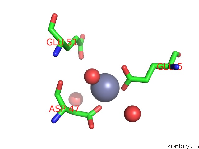
Mono view
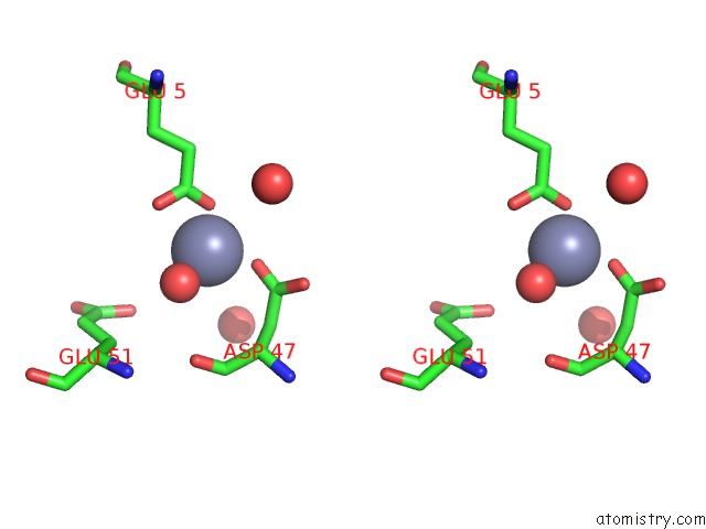
Stereo pair view
Mono view
Stereo pair view
A full contact list of Zinc with other atoms in the Zn binding
site number 1 of X-Ray Structure of Calcium-Loaded Calbindomodulin (A Calbindin D9K Re- Engineered to Undergo A Conformational Opening) at 1.44 A Resolution within 5.0Å range:
|
Zinc binding site 2 out of 2 in 1qx2
Go back to
Zinc binding site 2 out
of 2 in the X-Ray Structure of Calcium-Loaded Calbindomodulin (A Calbindin D9K Re- Engineered to Undergo A Conformational Opening) at 1.44 A Resolution
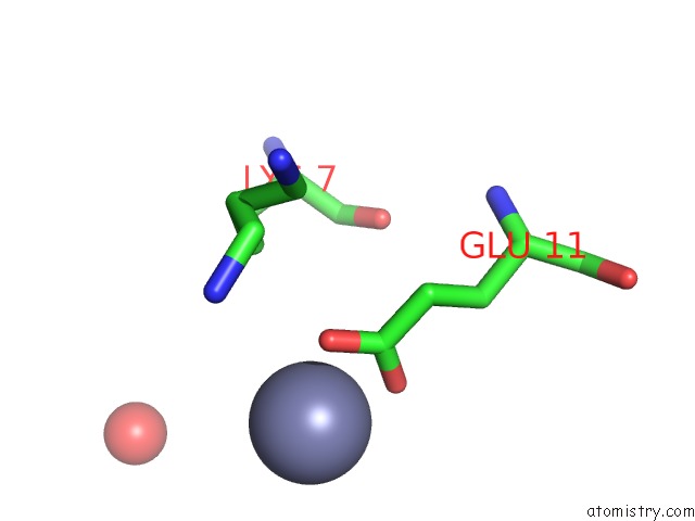
Mono view
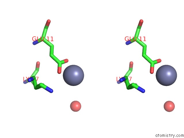
Stereo pair view
Mono view
Stereo pair view
A full contact list of Zinc with other atoms in the Zn binding
site number 2 of X-Ray Structure of Calcium-Loaded Calbindomodulin (A Calbindin D9K Re- Engineered to Undergo A Conformational Opening) at 1.44 A Resolution within 5.0Å range:
|
Reference:
C.G.Bunick,
M.R.Nelson,
S.Mangahas,
M.J.Hunter,
J.H.Sheehan,
L.S.Mizoue,
G.J.Bunick,
W.J.Chazin.
Designing Sequence to Control Protein Function in An Ef-Hand Protein J.Am.Chem.Soc. V. 126 5990 2004.
ISSN: ISSN 0002-7863
PubMed: 15137763
DOI: 10.1021/JA0397456
Page generated: Wed Oct 16 18:18:02 2024
ISSN: ISSN 0002-7863
PubMed: 15137763
DOI: 10.1021/JA0397456
Last articles
K in 5U41K in 5U3Q
K in 5U3G
K in 5TZO
K in 5U06
K in 5TXG
K in 5TVV
K in 5TXR
K in 5TOY
K in 5TOP