Zinc »
PDB 1qmu-1r23 »
1qqt »
Zinc in PDB 1qqt: Methionyl-Trna Synthetase From Escherichia Coli
Enzymatic activity of Methionyl-Trna Synthetase From Escherichia Coli
All present enzymatic activity of Methionyl-Trna Synthetase From Escherichia Coli:
6.1.1.10;
6.1.1.10;
Protein crystallography data
The structure of Methionyl-Trna Synthetase From Escherichia Coli, PDB code: 1qqt
was solved by
Y.Mechulam,
E.Schmitt,
L.Maveyraud,
C.Zelwer,
O.Nureki,
S.Yokoyama,
M.Konno,
S.Blanquet,
with X-Ray Crystallography technique. A brief refinement statistics is given in the table below:
| Resolution Low / High (Å) | 20.00 / 2.03 |
| Space group | P 1 21 1 |
| Cell size a, b, c (Å), α, β, γ (°) | 78.150, 46.300, 87.700, 90.00, 109.06, 90.00 |
| R / Rfree (%) | 17 / 21.4 |
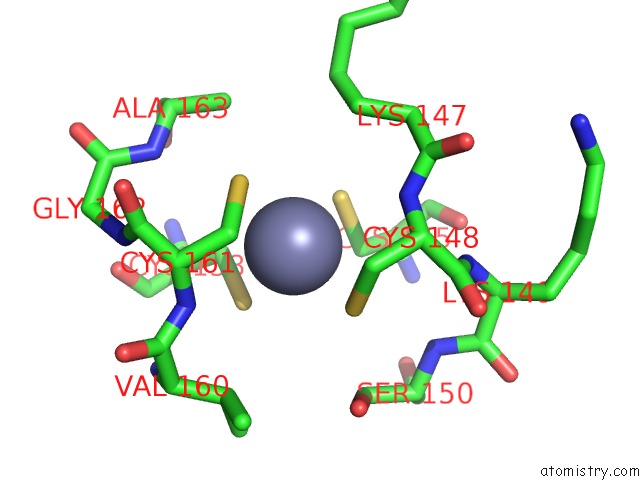
Zinc Binding Sites:
The binding sites of Zinc atom in the Methionyl-Trna Synthetase From Escherichia Coli
(pdb code 1qqt). This binding sites where shown within
5.0 Angstroms radius around Zinc atom.
In total only one binding site of Zinc was determined in the Methionyl-Trna Synthetase From Escherichia Coli, PDB code: 1qqt:
In total only one binding site of Zinc was determined in the Methionyl-Trna Synthetase From Escherichia Coli, PDB code: 1qqt:
Zinc binding site 1 out of 1 in 1qqt
Go back to
Zinc binding site 1 out
of 1 in the Methionyl-Trna Synthetase From Escherichia Coli
Mono view
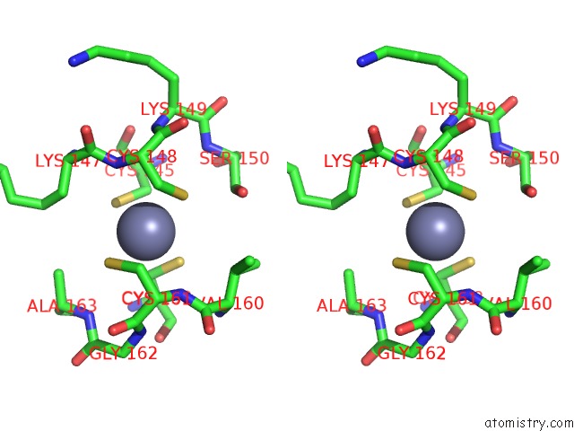
Stereo pair view
Mono view
Stereo pair view
A full contact list of Zinc with other atoms in the Zn binding
site number 1 of Methionyl-Trna Synthetase From Escherichia Coli within 5.0Å range:
|
Reference:
Y.Mechulam,
E.Schmitt,
L.Maveyraud,
C.Zelwer,
O.Nureki,
S.Yokoyama,
M.Konno,
S.Blanquet.
Crystal Structure of Escherichia Coli Methionyl-Trna Synthetase Highlights Species-Specific Features. J.Mol.Biol. V. 294 1287 1999.
ISSN: ISSN 0022-2836
PubMed: 10600385
DOI: 10.1006/JMBI.1999.3339
Page generated: Wed Oct 16 18:14:35 2024
ISSN: ISSN 0022-2836
PubMed: 10600385
DOI: 10.1006/JMBI.1999.3339
Last articles
Mg in 5BTLMg in 5BTM
Mg in 5BTI
Mg in 5BTG
Mg in 5BTF
Mg in 5BTD
Mg in 5BTC
Mg in 5BTA
Mg in 5BON
Mg in 5BSU