Zinc »
PDB 1q66-1qmd »
1qiy »
Zinc in PDB 1qiy: Human Insulin Hexamers with Chain B His Mutated to Tyr Complexed with Phenol
Protein crystallography data
The structure of Human Insulin Hexamers with Chain B His Mutated to Tyr Complexed with Phenol, PDB code: 1qiy
was solved by
L.Tang,
J.L.Whittingham,
C.S.Verma,
L.S.D.Caves,
G.G.Dodson,
with X-Ray Crystallography technique. A brief refinement statistics is given in the table below:
| Resolution Low / High (Å) | 36.80 / 2.30 |
| Space group | P 1 21 1 |
| Cell size a, b, c (Å), α, β, γ (°) | 61.100, 62.080, 48.350, 90.00, 109.87, 90.00 |
| R / Rfree (%) | 18.6 / n/a |
Other elements in 1qiy:
The structure of Human Insulin Hexamers with Chain B His Mutated to Tyr Complexed with Phenol also contains other interesting chemical elements:
| Chlorine | (Cl) | 2 atoms |
Zinc Binding Sites:
The binding sites of Zinc atom in the Human Insulin Hexamers with Chain B His Mutated to Tyr Complexed with Phenol
(pdb code 1qiy). This binding sites where shown within
5.0 Angstroms radius around Zinc atom.
In total 2 binding sites of Zinc where determined in the Human Insulin Hexamers with Chain B His Mutated to Tyr Complexed with Phenol, PDB code: 1qiy:
Jump to Zinc binding site number: 1; 2;
In total 2 binding sites of Zinc where determined in the Human Insulin Hexamers with Chain B His Mutated to Tyr Complexed with Phenol, PDB code: 1qiy:
Jump to Zinc binding site number: 1; 2;
Zinc binding site 1 out of 2 in 1qiy
Go back to
Zinc binding site 1 out
of 2 in the Human Insulin Hexamers with Chain B His Mutated to Tyr Complexed with Phenol
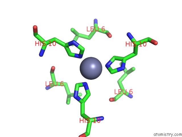
Mono view
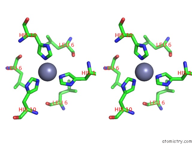
Stereo pair view
Mono view
Stereo pair view
A full contact list of Zinc with other atoms in the Zn binding
site number 1 of Human Insulin Hexamers with Chain B His Mutated to Tyr Complexed with Phenol within 5.0Å range:
|
Zinc binding site 2 out of 2 in 1qiy
Go back to
Zinc binding site 2 out
of 2 in the Human Insulin Hexamers with Chain B His Mutated to Tyr Complexed with Phenol
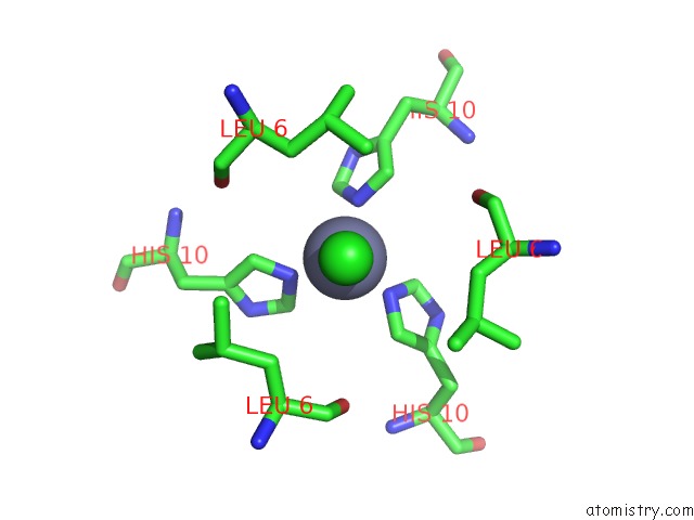
Mono view
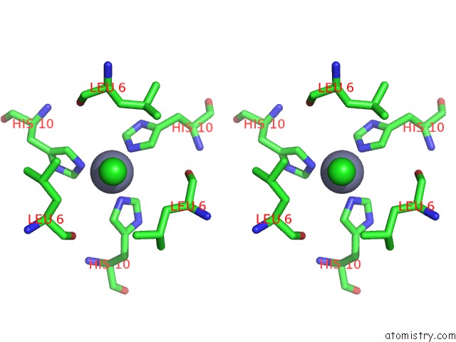
Stereo pair view
Mono view
Stereo pair view
A full contact list of Zinc with other atoms in the Zn binding
site number 2 of Human Insulin Hexamers with Chain B His Mutated to Tyr Complexed with Phenol within 5.0Å range:
|
Reference:
L.Tang,
J.L.Whittingham,
C.S.Verma,
L.S.D.Caves,
G.G.Dodson.
Structural Consequences of the B5 Histidine --> Tyrosine Mutation in Human Insulin Characterized By X-Ray Crystallography and Conformational Analysis. Biochemistry V. 38 12041 1999.
ISSN: ISSN 0006-2960
PubMed: 10508408
DOI: 10.1021/BI990700K
Page generated: Wed Oct 16 18:11:24 2024
ISSN: ISSN 0006-2960
PubMed: 10508408
DOI: 10.1021/BI990700K
Last articles
K in 5SCLK in 5T5M
K in 5T30
K in 5T09
K in 5SHY
K in 5SCK
K in 5SCJ
K in 5SCI
K in 5SCH
K in 5SCG