Zinc »
PDB 1ndz-1nve »
1nnj »
Zinc in PDB 1nnj: Crystal Structure Complex Between the Lactococcus Lactis Fpg and An Abasic Site Containing Dna
Enzymatic activity of Crystal Structure Complex Between the Lactococcus Lactis Fpg and An Abasic Site Containing Dna
All present enzymatic activity of Crystal Structure Complex Between the Lactococcus Lactis Fpg and An Abasic Site Containing Dna:
3.2.2.23;
3.2.2.23;
Protein crystallography data
The structure of Crystal Structure Complex Between the Lactococcus Lactis Fpg and An Abasic Site Containing Dna, PDB code: 1nnj
was solved by
L.Serre,
K.Pereira De Jesus,
S.Boiteux,
C.Zelwer,
B.Castaing,
with X-Ray Crystallography technique. A brief refinement statistics is given in the table below:
| Resolution Low / High (Å) | 35.00 / 1.90 |
| Space group | P 41 21 2 |
| Cell size a, b, c (Å), α, β, γ (°) | 92.150, 92.150, 142.530, 90.00, 90.00, 90.00 |
| R / Rfree (%) | 21.5 / 23.6 |
Zinc Binding Sites:
The binding sites of Zinc atom in the Crystal Structure Complex Between the Lactococcus Lactis Fpg and An Abasic Site Containing Dna
(pdb code 1nnj). This binding sites where shown within
5.0 Angstroms radius around Zinc atom.
In total only one binding site of Zinc was determined in the Crystal Structure Complex Between the Lactococcus Lactis Fpg and An Abasic Site Containing Dna, PDB code: 1nnj:
In total only one binding site of Zinc was determined in the Crystal Structure Complex Between the Lactococcus Lactis Fpg and An Abasic Site Containing Dna, PDB code: 1nnj:
Zinc binding site 1 out of 1 in 1nnj
Go back to
Zinc binding site 1 out
of 1 in the Crystal Structure Complex Between the Lactococcus Lactis Fpg and An Abasic Site Containing Dna
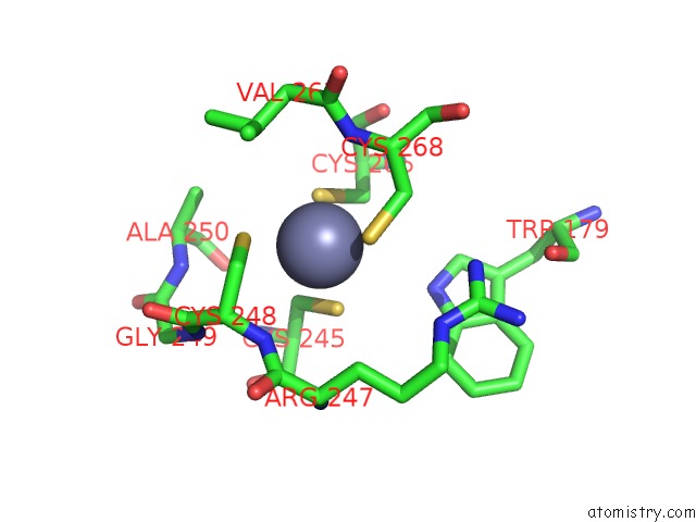
Mono view
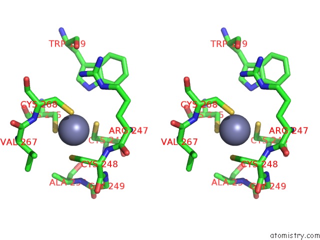
Stereo pair view
Mono view
Stereo pair view
A full contact list of Zinc with other atoms in the Zn binding
site number 1 of Crystal Structure Complex Between the Lactococcus Lactis Fpg and An Abasic Site Containing Dna within 5.0Å range:
|
Reference:
K.Pereira De Jesus,
L.Serre,
C.Zelwer,
B.Castaing.
Structural Insights Into Abasic Site For Fpg Specific Binding and Catalysis: Comparative High-Resolution Crystallographic Studies of Fpg Bound to Various Models of Abasic Site Analogues-Containing Dna. Nucleic Acids Res. V. 33 5936 2005.
ISSN: ISSN 0305-1048
PubMed: 16243784
DOI: 10.1093/NAR/GKI879
Page generated: Wed Oct 16 17:19:16 2024
ISSN: ISSN 0305-1048
PubMed: 16243784
DOI: 10.1093/NAR/GKI879
Last articles
Mg in 1MCZMg in 1MEZ
Mg in 1MDR
Mg in 1MDL
Mg in 1MC1
Mg in 1MC3
Mg in 1MBZ
Mg in 1MB9
Mg in 1MB3
Mg in 1MAU