Atomistry » Zinc » PDB 1ndz-1nve » 1nfg
Atomistry »
  Zinc »
    PDB 1ndz-1nve »
      1nfg »

Zinc in PDB 1nfg: Structure of D-Hydantoinase

Enzymatic activity of Structure of D-Hydantoinase

All present enzymatic activity of Structure of D-Hydantoinase:
3.5.2.2;

Protein crystallography data

The structure of Structure of D-Hydantoinase, PDB code: 1nfg was solved by Z.Xu, Y.Yang, W.Jiang, E.Arnold, J.Ding, with X-Ray Crystallography technique. A brief refinement statistics is given in the table below:

Resolution Low / High (Å) 14.94 / 2.70
Space group P 1 21 1
Cell size a, b, c (Å), α, β, γ (°) 68.700, 170.500, 87.300, 90.00, 95.20, 90.00
R / Rfree (%) 22 / 23.9

Zinc Binding Sites:

The binding sites of Zinc atom in the Structure of D-Hydantoinase (pdb code 1nfg). This binding sites where shown within 5.0 Angstroms radius around Zinc atom.
In total 8 binding sites of Zinc where determined in the Structure of D-Hydantoinase, PDB code: 1nfg:
Jump to Zinc binding site number: 1; 2; 3; 4; 5; 6; 7; 8;

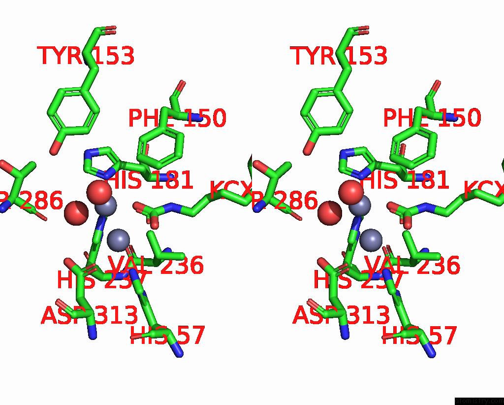
Zinc binding site 1 out of 8 in 1nfg

Go back to Zinc Binding Sites List in 1nfg
Zinc binding site 1 out of 8 in the Structure of D-Hydantoinase


Mono view


Stereo pair view

A full contact list of Zinc with other atoms in the Zn binding site number 1 of Structure of D-Hydantoinase within 5.0Å range:
probe atom residue distance (Å) B Occ
A:Zn501

b:21.0
occ:1.00
OD1 A:ASP313 2.2 25.2 1.0
NE2 A:HIS59 2.3 31.3 1.0
NE2 A:HIS57 2.4 26.4 1.0
OQ2 A:KCX148 2.5 12.4 1.0
CG A:ASP313 3.1 27.5 1.0
ZN A:ZN502 3.2 22.7 1.0
CX A:KCX148 3.2 17.9 1.0
OQ1 A:KCX148 3.2 13.7 1.0
CD2 A:HIS59 3.3 29.6 1.0
O A:HOH579 3.3 24.0 1.0
CE1 A:HIS57 3.3 25.5 1.0
CE1 A:HIS59 3.3 29.5 1.0
O A:HOH581 3.3 13.7 1.0
CD2 A:HIS57 3.4 25.5 1.0
OD2 A:ASP313 3.6 29.6 1.0
CB A:ASP313 4.4 25.6 1.0
ND1 A:HIS59 4.4 28.8 1.0
CG A:HIS59 4.4 29.6 1.0
ND1 A:HIS57 4.4 24.9 1.0
NZ A:KCX148 4.5 14.6 1.0
CG A:HIS57 4.5 24.4 1.0
CD2 A:HIS237 4.6 27.0 1.0
CA A:ASP313 4.8 25.5 1.0
CE2 A:PHE91 4.8 21.4 1.0
NE2 A:HIS237 4.8 26.8 1.0
CD2 A:PHE91 4.9 23.3 1.0
O A:HOH580 5.0 21.0 1.0

Zinc binding site 2 out of 8 in 1nfg

Go back to Zinc Binding Sites List in 1nfg
Zinc binding site 2 out of 8 in the Structure of D-Hydantoinase


Mono view


Stereo pair view

A full contact list of Zinc with other atoms in the Zn binding site number 2 of Structure of D-Hydantoinase within 5.0Å range:
probe atom residue distance (Å) B Occ
A:Zn502

b:22.7
occ:1.00
OQ1 A:KCX148 2.0 13.7 1.0
O A:HOH579 2.2 24.0 1.0
ND1 A:HIS181 2.4 21.8 1.0
NE2 A:HIS237 2.6 26.8 1.0
CE1 A:HIS181 3.1 20.9 1.0
CX A:KCX148 3.1 17.9 1.0
ZN A:ZN501 3.2 21.0 1.0
CD2 A:HIS237 3.2 27.0 1.0
O A:HOH581 3.4 13.7 1.0
CG A:HIS181 3.6 20.3 1.0
OQ2 A:KCX148 3.7 12.4 1.0
CE1 A:HIS237 3.8 26.6 1.0
CE2 A:PHE150 4.0 22.6 1.0
CB A:HIS181 4.1 21.4 1.0
O A:THR286 4.2 21.6 1.0
NZ A:KCX148 4.2 14.6 1.0
NE2 A:HIS181 4.3 20.5 1.0
OD2 A:ASP313 4.4 29.6 1.0
CG A:HIS237 4.5 27.4 1.0
CD2 A:HIS181 4.6 21.9 1.0
OD1 A:ASP313 4.6 25.2 1.0
CG2 A:VAL236 4.7 19.1 1.0
NE2 A:HIS57 4.7 26.4 1.0
CZ A:PHE150 4.7 21.9 1.0
ND1 A:HIS237 4.7 28.3 1.0
CE1 A:HIS57 4.8 25.5 1.0
CD2 A:PHE150 4.8 22.6 1.0
OH A:TYR153 4.8 44.0 1.0
CG A:ASP313 4.9 27.5 1.0
CA A:HIS181 5.0 21.7 1.0

Zinc binding site 3 out of 8 in 1nfg

Go back to Zinc Binding Sites List in 1nfg
Zinc binding site 3 out of 8 in the Structure of D-Hydantoinase


Mono view


Stereo pair view

A full contact list of Zinc with other atoms in the Zn binding site number 3 of Structure of D-Hydantoinase within 5.0Å range:
probe atom residue distance (Å) B Occ
B:Zn601

b:21.0
occ:1.00
OD1 B:ASP313 2.2 24.9 1.0
NE2 B:HIS59 2.3 31.0 1.0
NE2 B:HIS57 2.4 26.1 1.0
OQ2 B:KCX148 2.5 12.2 1.0
CG B:ASP313 3.1 27.2 1.0
ZN B:ZN602 3.2 22.7 1.0
CX B:KCX148 3.2 17.6 1.0
OQ1 B:KCX148 3.2 13.5 1.0
CD2 B:HIS59 3.3 29.3 1.0
O B:HOH689 3.3 24.0 1.0
CE1 B:HIS57 3.3 25.2 1.0
CE1 B:HIS59 3.3 29.2 1.0
O B:HOH691 3.3 13.7 1.0
CD2 B:HIS57 3.4 25.2 1.0
OD2 B:ASP313 3.6 29.4 1.0
CB B:ASP313 4.4 25.4 1.0
ND1 B:HIS59 4.4 28.5 1.0
CG B:HIS59 4.4 29.4 1.0
ND1 B:HIS57 4.4 24.6 1.0
NZ B:KCX148 4.5 14.3 1.0
CG B:HIS57 4.5 24.2 1.0
CD2 B:HIS237 4.6 26.8 1.0
CA B:ASP313 4.8 25.2 1.0
CE2 B:PHE91 4.8 21.1 1.0
NE2 B:HIS237 4.8 26.5 1.0
CD2 B:PHE91 4.9 23.0 1.0
O B:HOH690 5.0 21.0 1.0

Zinc binding site 4 out of 8 in 1nfg

Go back to Zinc Binding Sites List in 1nfg
Zinc binding site 4 out of 8 in the Structure of D-Hydantoinase


Mono view


Stereo pair view

A full contact list of Zinc with other atoms in the Zn binding site number 4 of Structure of D-Hydantoinase within 5.0Å range:
probe atom residue distance (Å) B Occ
B:Zn602

b:22.7
occ:1.00
OQ1 B:KCX148 2.0 13.5 1.0
O B:HOH689 2.2 24.0 1.0
ND1 B:HIS181 2.4 21.5 1.0
NE2 B:HIS237 2.6 26.5 1.0
CE1 B:HIS181 3.1 20.7 1.0
CX B:KCX148 3.1 17.6 1.0
ZN B:ZN601 3.2 21.0 1.0
CD2 B:HIS237 3.2 26.8 1.0
O B:HOH691 3.4 13.7 1.0
CG B:HIS181 3.6 20.1 1.0
OQ2 B:KCX148 3.7 12.2 1.0
CE1 B:HIS237 3.8 26.4 1.0
CE2 B:PHE150 4.0 22.4 1.0
CB B:HIS181 4.1 21.2 1.0
O B:THR286 4.2 21.4 1.0
NZ B:KCX148 4.2 14.3 1.0
NE2 B:HIS181 4.3 20.2 1.0
OD2 B:ASP313 4.4 29.4 1.0
CG B:HIS237 4.5 27.1 1.0
CD2 B:HIS181 4.6 21.6 1.0
OD1 B:ASP313 4.6 24.9 1.0
CG2 B:VAL236 4.7 18.8 1.0
NE2 B:HIS57 4.7 26.1 1.0
CZ B:PHE150 4.7 21.6 1.0
ND1 B:HIS237 4.7 28.0 1.0
CE1 B:HIS57 4.8 25.2 1.0
CD2 B:PHE150 4.8 22.4 1.0
OH B:TYR153 4.8 43.8 1.0
CG B:ASP313 4.9 27.2 1.0
CA B:HIS181 5.0 21.4 1.0

Zinc binding site 5 out of 8 in 1nfg

Go back to Zinc Binding Sites List in 1nfg
Zinc binding site 5 out of 8 in the Structure of D-Hydantoinase


Mono view


Stereo pair view

A full contact list of Zinc with other atoms in the Zn binding site number 5 of Structure of D-Hydantoinase within 5.0Å range:
probe atom residue distance (Å) B Occ
C:Zn701

b:21.0
occ:1.00
OD1 C:ASP313 2.2 24.9 1.0
NE2 C:HIS59 2.3 31.0 1.0
NE2 C:HIS57 2.4 26.1 1.0
OQ2 C:KCX148 2.5 12.2 1.0
CG C:ASP313 3.1 27.2 1.0
ZN C:ZN702 3.2 22.7 1.0
CX C:KCX148 3.2 17.6 1.0
OQ1 C:KCX148 3.2 13.5 1.0
CD2 C:HIS59 3.3 29.3 1.0
O C:HOH782 3.3 24.0 1.0
CE1 C:HIS57 3.3 25.2 1.0
CE1 C:HIS59 3.3 29.2 1.0
O C:HOH784 3.3 13.7 1.0
CD2 C:HIS57 3.4 25.2 1.0
OD2 C:ASP313 3.6 29.4 1.0
CB C:ASP313 4.4 25.4 1.0
ND1 C:HIS59 4.4 28.5 1.0
CG C:HIS59 4.4 29.4 1.0
ND1 C:HIS57 4.4 24.6 1.0
NZ C:KCX148 4.5 14.3 1.0
CG C:HIS57 4.5 24.2 1.0
CD2 C:HIS237 4.6 26.8 1.0
CA C:ASP313 4.8 25.2 1.0
CE2 C:PHE91 4.8 21.1 1.0
NE2 C:HIS237 4.8 26.5 1.0
CD2 C:PHE91 4.9 23.0 1.0
O C:HOH783 5.0 21.0 1.0

Zinc binding site 6 out of 8 in 1nfg

Go back to Zinc Binding Sites List in 1nfg
Zinc binding site 6 out of 8 in the Structure of D-Hydantoinase


Mono view


Stereo pair view

A full contact list of Zinc with other atoms in the Zn binding site number 6 of Structure of D-Hydantoinase within 5.0Å range:
probe atom residue distance (Å) B Occ
C:Zn702

b:22.7
occ:1.00
OQ1 C:KCX148 2.0 13.5 1.0
O C:HOH782 2.2 24.0 1.0
ND1 C:HIS181 2.4 21.5 1.0
NE2 C:HIS237 2.6 26.5 1.0
CE1 C:HIS181 3.1 20.7 1.0
CX C:KCX148 3.1 17.6 1.0
ZN C:ZN701 3.2 21.0 1.0
CD2 C:HIS237 3.2 26.8 1.0
O C:HOH784 3.4 13.7 1.0
CG C:HIS181 3.6 20.1 1.0
OQ2 C:KCX148 3.7 12.2 1.0
CE1 C:HIS237 3.8 26.4 1.0
CE2 C:PHE150 4.0 22.4 1.0
CB C:HIS181 4.1 21.2 1.0
O C:THR286 4.2 21.4 1.0
NZ C:KCX148 4.2 14.3 1.0
NE2 C:HIS181 4.3 20.2 1.0
OD2 C:ASP313 4.4 29.4 1.0
CG C:HIS237 4.5 27.1 1.0
CD2 C:HIS181 4.6 21.6 1.0
OD1 C:ASP313 4.6 24.9 1.0
CG2 C:VAL236 4.7 18.8 1.0
NE2 C:HIS57 4.7 26.1 1.0
CZ C:PHE150 4.7 21.6 1.0
ND1 C:HIS237 4.7 28.0 1.0
CE1 C:HIS57 4.8 25.2 1.0
CD2 C:PHE150 4.8 22.4 1.0
OH C:TYR153 4.8 43.8 1.0
CG C:ASP313 4.9 27.2 1.0
CA C:HIS181 5.0 21.4 1.0

Zinc binding site 7 out of 8 in 1nfg

Go back to Zinc Binding Sites List in 1nfg
Zinc binding site 7 out of 8 in the Structure of D-Hydantoinase


Mono view


Stereo pair view

A full contact list of Zinc with other atoms in the Zn binding site number 7 of Structure of D-Hydantoinase within 5.0Å range:
probe atom residue distance (Å) B Occ
D:Zn801

b:21.0
occ:1.00
OD1 D:ASP313 2.2 24.9 1.0
NE2 D:HIS59 2.3 31.0 1.0
NE2 D:HIS57 2.4 26.1 1.0
OQ2 D:KCX148 2.5 12.2 1.0
CG D:ASP313 3.1 27.2 1.0
ZN D:ZN802 3.2 22.7 1.0
CX D:KCX148 3.2 17.6 1.0
OQ1 D:KCX148 3.2 13.5 1.0
CD2 D:HIS59 3.3 29.3 1.0
O D:HOH3085 3.3 24.0 1.0
CE1 D:HIS57 3.3 25.2 1.0
CE1 D:HIS59 3.3 29.2 1.0
O D:HOH3087 3.3 13.7 1.0
CD2 D:HIS57 3.4 25.2 1.0
OD2 D:ASP313 3.6 29.4 1.0
CB D:ASP313 4.4 25.4 1.0
ND1 D:HIS59 4.4 28.5 1.0
CG D:HIS59 4.4 29.4 1.0
ND1 D:HIS57 4.4 24.6 1.0
NZ D:KCX148 4.5 14.3 1.0
CG D:HIS57 4.5 24.2 1.0
CD2 D:HIS237 4.6 26.8 1.0
CA D:ASP313 4.8 25.2 1.0
CE2 D:PHE91 4.8 21.1 1.0
NE2 D:HIS237 4.8 26.5 1.0
CD2 D:PHE91 4.9 23.0 1.0
O D:HOH3086 5.0 21.0 1.0

Zinc binding site 8 out of 8 in 1nfg

Go back to Zinc Binding Sites List in 1nfg
Zinc binding site 8 out of 8 in the Structure of D-Hydantoinase


Mono view


Stereo pair view

A full contact list of Zinc with other atoms in the Zn binding site number 8 of Structure of D-Hydantoinase within 5.0Å range:
probe atom residue distance (Å) B Occ
D:Zn802

b:22.7
occ:1.00
OQ1 D:KCX148 2.0 13.5 1.0
O D:HOH3085 2.2 24.0 1.0
ND1 D:HIS181 2.4 21.5 1.0
NE2 D:HIS237 2.6 26.5 1.0
CE1 D:HIS181 3.1 20.7 1.0
CX D:KCX148 3.1 17.6 1.0
ZN D:ZN801 3.2 21.0 1.0
CD2 D:HIS237 3.2 26.8 1.0
O D:HOH3087 3.4 13.7 1.0
CG D:HIS181 3.6 20.1 1.0
OQ2 D:KCX148 3.7 12.2 1.0
CE1 D:HIS237 3.8 26.4 1.0
CE2 D:PHE150 4.0 22.4 1.0
CB D:HIS181 4.1 21.2 1.0
O D:THR286 4.2 21.4 1.0
NZ D:KCX148 4.2 14.3 1.0
NE2 D:HIS181 4.3 20.2 1.0
OD2 D:ASP313 4.4 29.4 1.0
CG D:HIS237 4.5 27.1 1.0
CD2 D:HIS181 4.6 21.6 1.0
OD1 D:ASP313 4.6 24.9 1.0
CG2 D:VAL236 4.7 18.8 1.0
NE2 D:HIS57 4.7 26.1 1.0
CZ D:PHE150 4.7 21.6 1.0
ND1 D:HIS237 4.7 28.0 1.0
CE1 D:HIS57 4.8 25.2 1.0
CD2 D:PHE150 4.8 22.4 1.0
OH D:TYR153 4.8 43.8 1.0
CG D:ASP313 4.9 27.2 1.0
CA D:HIS181 5.0 21.4 1.0

Reference:

Z.Xu, Y.Liu, Y.Yang, W.Jiang, E.Arnold, J.Ding. Crystal Structure of D-Hydantoinase From Burkholderia Pickettii at A Resolution of 2.7 Angstroms: Insights Into the Molecular Basis of Enzyme Thermostability. J.Bacteriol. V. 185 4038 2003.
ISSN: ISSN 0021-9193
PubMed: 12837777
DOI: 10.1128/JB.185.14.4038-4049.2003
Page generated: Wed Oct 16 17:15:44 2024

Last articles

Mg in 1SO2
Mg in 1SO5
Mg in 1SO4
Mg in 1SO3
Mg in 1SNF
Mg in 1SLH
Mg in 1SL2
Mg in 1SL5
Mg in 1SKR
Mg in 1SL0
© Copyright 2008-2020 by atomistry.com
Home   |    Site Map   |    Copyright   |    Contact us   |    Privacy