Zinc »
PDB 1my2-1ndy »
1nc8 »
Zinc in PDB 1nc8: High-Resolution Solution uc(Nmr) Structure of the Minimal Active Domain of the Human Immunodeficiency Virus Type-2 Nucleocapsid Protein, 15 Structures
Zinc Binding Sites:
The binding sites of Zinc atom in the High-Resolution Solution uc(Nmr) Structure of the Minimal Active Domain of the Human Immunodeficiency Virus Type-2 Nucleocapsid Protein, 15 Structures
(pdb code 1nc8). This binding sites where shown within
5.0 Angstroms radius around Zinc atom.
In total only one binding site of Zinc was determined in the High-Resolution Solution uc(Nmr) Structure of the Minimal Active Domain of the Human Immunodeficiency Virus Type-2 Nucleocapsid Protein, 15 Structures, PDB code: 1nc8:
In total only one binding site of Zinc was determined in the High-Resolution Solution uc(Nmr) Structure of the Minimal Active Domain of the Human Immunodeficiency Virus Type-2 Nucleocapsid Protein, 15 Structures, PDB code: 1nc8:
Zinc binding site 1 out of 1 in 1nc8
Go back to
Zinc binding site 1 out
of 1 in the High-Resolution Solution uc(Nmr) Structure of the Minimal Active Domain of the Human Immunodeficiency Virus Type-2 Nucleocapsid Protein, 15 Structures
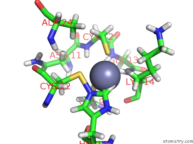
Mono view
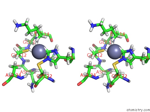
Stereo pair view
Mono view
Stereo pair view
A full contact list of Zinc with other atoms in the Zn binding
site number 1 of High-Resolution Solution uc(Nmr) Structure of the Minimal Active Domain of the Human Immunodeficiency Virus Type-2 Nucleocapsid Protein, 15 Structures within 5.0Å range:
|
Reference:
Y.Kodera,
K.Sato,
T.Tsukahara,
H.Komatsu,
T.Maeda,
T.Kohno.
High-Resolution Solution uc(Nmr) Structure of the Minimal Active Domain of the Human Immunodeficiency Virus Type-2 Nucleocapsid Protein. Biochemistry V. 37 17704 1998.
ISSN: ISSN 0006-2960
PubMed: 9922136
DOI: 10.1021/BI981818O
Page generated: Wed Oct 16 17:14:22 2024
ISSN: ISSN 0006-2960
PubMed: 9922136
DOI: 10.1021/BI981818O
Last articles
Na in 2JH6Na in 2JH5
Na in 2JH0
Na in 2J6L
Na in 2JGP
Na in 2JCA
Na in 2JBW
Na in 2JC4
Na in 2JBY
Na in 2JBA