Zinc »
PDB 1kzy-1lg6 »
1l7o »
Zinc in PDB 1l7o: Crystal Structure of Phosphoserine Phosphatase in Apo Form
Enzymatic activity of Crystal Structure of Phosphoserine Phosphatase in Apo Form
All present enzymatic activity of Crystal Structure of Phosphoserine Phosphatase in Apo Form:
3.1.3.3;
3.1.3.3;
Protein crystallography data
The structure of Crystal Structure of Phosphoserine Phosphatase in Apo Form, PDB code: 1l7o
was solved by
W.Wang,
H.S.Cho,
R.Kim,
J.Jancarik,
H.Yokota,
H.H.Nguyen,
I.V.Grigoriev,
D.E.Wemmer,
S.H.Kim,
Berkeley Structural Genomics Center (Bsgc),
with X-Ray Crystallography technique. A brief refinement statistics is given in the table below:
| Resolution Low / High (Å) | 15.00 / 2.20 |
| Space group | P 21 21 21 |
| Cell size a, b, c (Å), α, β, γ (°) | 36.888, 117.889, 117.361, 90.00, 90.00, 90.00 |
| R / Rfree (%) | 22.4 / 25.4 |
Zinc Binding Sites:
The binding sites of Zinc atom in the Crystal Structure of Phosphoserine Phosphatase in Apo Form
(pdb code 1l7o). This binding sites where shown within
5.0 Angstroms radius around Zinc atom.
In total 3 binding sites of Zinc where determined in the Crystal Structure of Phosphoserine Phosphatase in Apo Form, PDB code: 1l7o:
Jump to Zinc binding site number: 1; 2; 3;
In total 3 binding sites of Zinc where determined in the Crystal Structure of Phosphoserine Phosphatase in Apo Form, PDB code: 1l7o:
Jump to Zinc binding site number: 1; 2; 3;
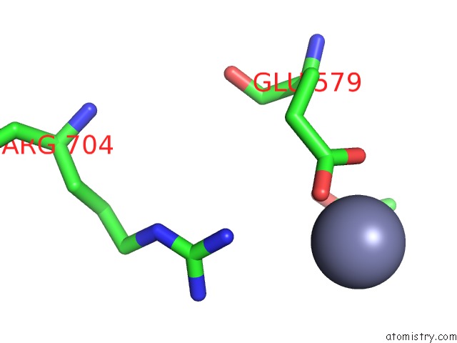
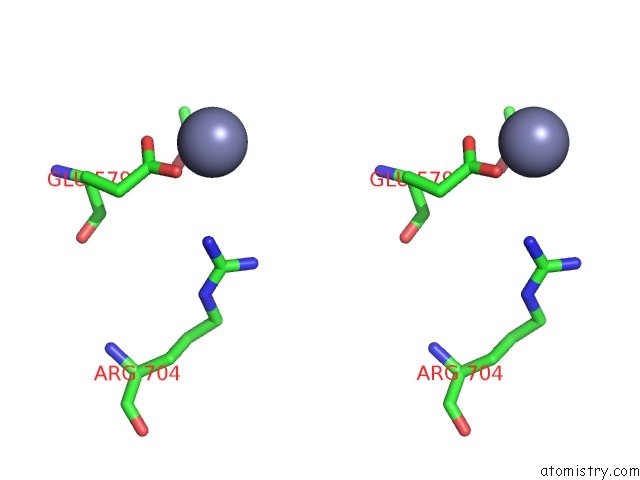
Zinc binding site 1 out of 3 in 1l7o
Go back to
Zinc binding site 1 out
of 3 in the Crystal Structure of Phosphoserine Phosphatase in Apo Form
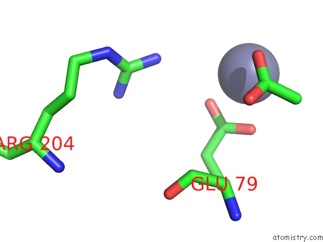
Mono view
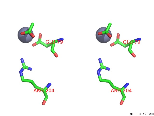
Stereo pair view
Mono view
Stereo pair view
A full contact list of Zinc with other atoms in the Zn binding
site number 1 of Crystal Structure of Phosphoserine Phosphatase in Apo Form within 5.0Å range:
|
Zinc binding site 2 out of 3 in 1l7o
Go back to
Zinc binding site 2 out
of 3 in the Crystal Structure of Phosphoserine Phosphatase in Apo Form
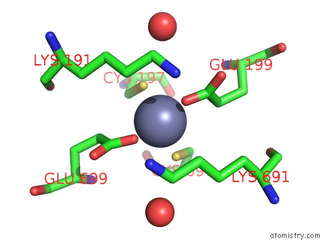
Mono view
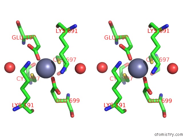
Stereo pair view
Mono view
Stereo pair view
A full contact list of Zinc with other atoms in the Zn binding
site number 2 of Crystal Structure of Phosphoserine Phosphatase in Apo Form within 5.0Å range:
|
Zinc binding site 3 out of 3 in 1l7o
Go back to
Zinc binding site 3 out
of 3 in the Crystal Structure of Phosphoserine Phosphatase in Apo Form
Mono view
Stereo pair view
Mono view
Stereo pair view
A full contact list of Zinc with other atoms in the Zn binding
site number 3 of Crystal Structure of Phosphoserine Phosphatase in Apo Form within 5.0Å range:
|
Reference:
W.Wang,
H.S.Cho,
R.Kim,
J.Jancarik,
H.Yokota,
H.H.Nguyen,
I.V.Grigoriev,
D.E.Wemmer,
S.H.Kim.
Structural Characterization of the Reaction Pathway in Phosphoserine Phosphatase: Crystallographic "Snapshots" of Intermediate States. J.Mol.Biol. V. 319 421 2002.
ISSN: ISSN 0022-2836
PubMed: 12051918
DOI: 10.1016/S0022-2836(02)00324-8
Page generated: Sun Oct 13 04:50:29 2024
ISSN: ISSN 0022-2836
PubMed: 12051918
DOI: 10.1016/S0022-2836(02)00324-8
Last articles
Fe in 2YXOFe in 2YRS
Fe in 2YXC
Fe in 2YNM
Fe in 2YVJ
Fe in 2YP1
Fe in 2YU2
Fe in 2YU1
Fe in 2YQB
Fe in 2YOO