Zinc »
PDB 1kk1-1kzp »
1kys »
Zinc in PDB 1kys: Crystal Structure of A Zn-Bound Green Fluorescent Protein Biosensor
Protein crystallography data
The structure of Crystal Structure of A Zn-Bound Green Fluorescent Protein Biosensor, PDB code: 1kys
was solved by
D.P.Barondeau,
C.J.Kassmann,
J.A.Tainer,
E.D.Getzoff,
with X-Ray Crystallography technique. A brief refinement statistics is given in the table below:
| Resolution Low / High (Å) | 20.00 / 1.44 |
| Space group | P 21 21 21 |
| Cell size a, b, c (Å), α, β, γ (°) | 51.147, 62.226, 68.817, 90.00, 90.00, 90.00 |
| R / Rfree (%) | 15.7 / 22.9 |
Zinc Binding Sites:
The binding sites of Zinc atom in the Crystal Structure of A Zn-Bound Green Fluorescent Protein Biosensor
(pdb code 1kys). This binding sites where shown within
5.0 Angstroms radius around Zinc atom.
In total 4 binding sites of Zinc where determined in the Crystal Structure of A Zn-Bound Green Fluorescent Protein Biosensor, PDB code: 1kys:
Jump to Zinc binding site number: 1; 2; 3; 4;
In total 4 binding sites of Zinc where determined in the Crystal Structure of A Zn-Bound Green Fluorescent Protein Biosensor, PDB code: 1kys:
Jump to Zinc binding site number: 1; 2; 3; 4;
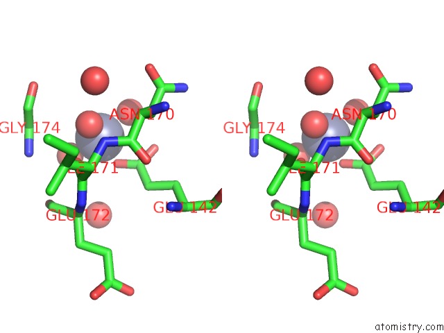
Zinc binding site 1 out of 4 in 1kys
Go back to
Zinc binding site 1 out
of 4 in the Crystal Structure of A Zn-Bound Green Fluorescent Protein Biosensor

Mono view

Stereo pair view
Mono view
Stereo pair view
A full contact list of Zinc with other atoms in the Zn binding
site number 1 of Crystal Structure of A Zn-Bound Green Fluorescent Protein Biosensor within 5.0Å range:
|
Zinc binding site 2 out of 4 in 1kys
Go back to
Zinc binding site 2 out
of 4 in the Crystal Structure of A Zn-Bound Green Fluorescent Protein Biosensor

Mono view
Stereo pair view
Mono view
Stereo pair view
A full contact list of Zinc with other atoms in the Zn binding
site number 2 of Crystal Structure of A Zn-Bound Green Fluorescent Protein Biosensor within 5.0Å range:
|
Zinc binding site 3 out of 4 in 1kys
Go back to
Zinc binding site 3 out
of 4 in the Crystal Structure of A Zn-Bound Green Fluorescent Protein Biosensor

Mono view

Stereo pair view
Mono view
Stereo pair view
A full contact list of Zinc with other atoms in the Zn binding
site number 3 of Crystal Structure of A Zn-Bound Green Fluorescent Protein Biosensor within 5.0Å range:
|
Zinc binding site 4 out of 4 in 1kys
Go back to
Zinc binding site 4 out
of 4 in the Crystal Structure of A Zn-Bound Green Fluorescent Protein Biosensor

Mono view

Stereo pair view
Mono view
Stereo pair view
A full contact list of Zinc with other atoms in the Zn binding
site number 4 of Crystal Structure of A Zn-Bound Green Fluorescent Protein Biosensor within 5.0Å range:
|
Reference:
D.P.Barondeau,
C.J.Kassmann,
J.A.Tainer,
E.D.Getzoff.
Structural Chemistry of A Green Fluorescent Protein Zn Biosensor. J.Am.Chem.Soc. V. 124 3522 2002.
ISSN: ISSN 0002-7863
PubMed: 11929238
DOI: 10.1021/JA0176954
Page generated: Sun Oct 13 04:42:49 2024
ISSN: ISSN 0002-7863
PubMed: 11929238
DOI: 10.1021/JA0176954
Last articles
Mg in 5I0DMg in 5I3U
Mg in 5I49
Mg in 5I2U
Mg in 5I2D
Mg in 5I1F
Mg in 5I0I
Mg in 5I0H
Mg in 5I10
Mg in 5HZF