Zinc »
PDB 1k6y-1kk0 »
1kap »
Zinc in PDB 1kap: Three-Dimensional Structure of the Alkaline Protease of Pseudomonas Aeruginosa: A Two-Domain Protein with A Calcium Binding Parallel Beta Roll Motif
Protein crystallography data
The structure of Three-Dimensional Structure of the Alkaline Protease of Pseudomonas Aeruginosa: A Two-Domain Protein with A Calcium Binding Parallel Beta Roll Motif, PDB code: 1kap
was solved by
U.Baumann,
S.Wu,
K.M.Flaherty,
D.B.Mckay,
with X-Ray Crystallography technique. A brief refinement statistics is given in the table below:
| Resolution Low / High (Å) | 10.00 / 1.64 |
| Space group | P 65 |
| Cell size a, b, c (Å), α, β, γ (°) | 106.900, 106.900, 97.000, 90.00, 90.00, 120.00 |
| R / Rfree (%) | 17.6 / n/a |
Other elements in 1kap:
The structure of Three-Dimensional Structure of the Alkaline Protease of Pseudomonas Aeruginosa: A Two-Domain Protein with A Calcium Binding Parallel Beta Roll Motif also contains other interesting chemical elements:
| Calcium | (Ca) | 8 atoms |
Zinc Binding Sites:
The binding sites of Zinc atom in the Three-Dimensional Structure of the Alkaline Protease of Pseudomonas Aeruginosa: A Two-Domain Protein with A Calcium Binding Parallel Beta Roll Motif
(pdb code 1kap). This binding sites where shown within
5.0 Angstroms radius around Zinc atom.
In total only one binding site of Zinc was determined in the Three-Dimensional Structure of the Alkaline Protease of Pseudomonas Aeruginosa: A Two-Domain Protein with A Calcium Binding Parallel Beta Roll Motif, PDB code: 1kap:
In total only one binding site of Zinc was determined in the Three-Dimensional Structure of the Alkaline Protease of Pseudomonas Aeruginosa: A Two-Domain Protein with A Calcium Binding Parallel Beta Roll Motif, PDB code: 1kap:
Zinc binding site 1 out of 1 in 1kap
Go back to
Zinc binding site 1 out
of 1 in the Three-Dimensional Structure of the Alkaline Protease of Pseudomonas Aeruginosa: A Two-Domain Protein with A Calcium Binding Parallel Beta Roll Motif
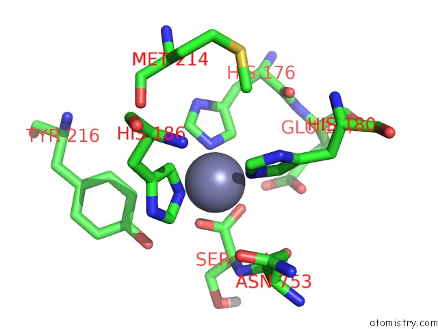
Mono view
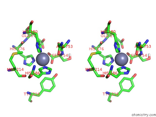
Stereo pair view
Mono view
Stereo pair view
A full contact list of Zinc with other atoms in the Zn binding
site number 1 of Three-Dimensional Structure of the Alkaline Protease of Pseudomonas Aeruginosa: A Two-Domain Protein with A Calcium Binding Parallel Beta Roll Motif within 5.0Å range:
|
Reference:
U.Baumann,
S.Wu,
K.M.Flaherty,
D.B.Mckay.
Three-Dimensional Structure of the Alkaline Protease of Pseudomonas Aeruginosa: A Two-Domain Protein with A Calcium Binding Parallel Beta Roll Motif. Embo J. V. 12 3357 1993.
ISSN: ISSN 0261-4189
PubMed: 8253063
Page generated: Sun Oct 13 04:12:13 2024
ISSN: ISSN 0261-4189
PubMed: 8253063
Last articles
Mg in 7BNRMg in 7BNK
Mg in 7BMC
Mg in 7BM9
Mg in 7BM8
Mg in 7BM6
Mg in 7BL4
Mg in 7BL6
Mg in 7BL5
Mg in 7BJT