Zinc »
PDB 1k6y-1kk0 »
1kae »
Zinc in PDB 1kae: L-Histidinol Dehydrogenase (Hisd) Structure Complexed with L- Histidinol (Substrate), Zinc and Nad (Cofactor)
Enzymatic activity of L-Histidinol Dehydrogenase (Hisd) Structure Complexed with L- Histidinol (Substrate), Zinc and Nad (Cofactor)
All present enzymatic activity of L-Histidinol Dehydrogenase (Hisd) Structure Complexed with L- Histidinol (Substrate), Zinc and Nad (Cofactor):
1.1.1.23;
1.1.1.23;
Protein crystallography data
The structure of L-Histidinol Dehydrogenase (Hisd) Structure Complexed with L- Histidinol (Substrate), Zinc and Nad (Cofactor), PDB code: 1kae
was solved by
J.A.R.G.Barbosa,
J.Sivaraman,
Y.Li,
R.Larocque,
A.Matte,
J.D.Schrag,
M.Cygler,
with X-Ray Crystallography technique. A brief refinement statistics is given in the table below:
| Resolution Low / High (Å) | 38.50 / 1.70 |
| Space group | P 21 21 21 |
| Cell size a, b, c (Å), α, β, γ (°) | 54.930, 107.940, 156.710, 90.00, 90.00, 90.00 |
| R / Rfree (%) | 21.3 / 24.1 |
Zinc Binding Sites:
The binding sites of Zinc atom in the L-Histidinol Dehydrogenase (Hisd) Structure Complexed with L- Histidinol (Substrate), Zinc and Nad (Cofactor)
(pdb code 1kae). This binding sites where shown within
5.0 Angstroms radius around Zinc atom.
In total 2 binding sites of Zinc where determined in the L-Histidinol Dehydrogenase (Hisd) Structure Complexed with L- Histidinol (Substrate), Zinc and Nad (Cofactor), PDB code: 1kae:
Jump to Zinc binding site number: 1; 2;
In total 2 binding sites of Zinc where determined in the L-Histidinol Dehydrogenase (Hisd) Structure Complexed with L- Histidinol (Substrate), Zinc and Nad (Cofactor), PDB code: 1kae:
Jump to Zinc binding site number: 1; 2;
Zinc binding site 1 out of 2 in 1kae
Go back to
Zinc binding site 1 out
of 2 in the L-Histidinol Dehydrogenase (Hisd) Structure Complexed with L- Histidinol (Substrate), Zinc and Nad (Cofactor)
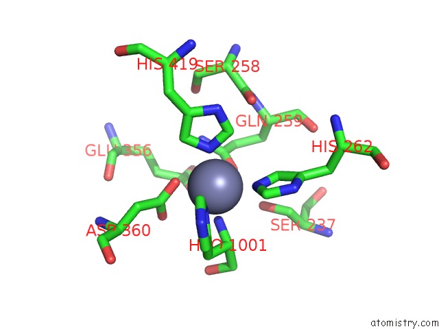
Mono view
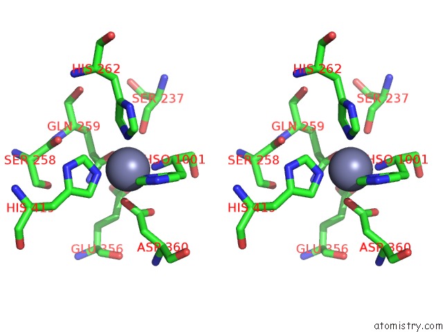
Stereo pair view
Mono view
Stereo pair view
A full contact list of Zinc with other atoms in the Zn binding
site number 1 of L-Histidinol Dehydrogenase (Hisd) Structure Complexed with L- Histidinol (Substrate), Zinc and Nad (Cofactor) within 5.0Å range:
|
Zinc binding site 2 out of 2 in 1kae
Go back to
Zinc binding site 2 out
of 2 in the L-Histidinol Dehydrogenase (Hisd) Structure Complexed with L- Histidinol (Substrate), Zinc and Nad (Cofactor)
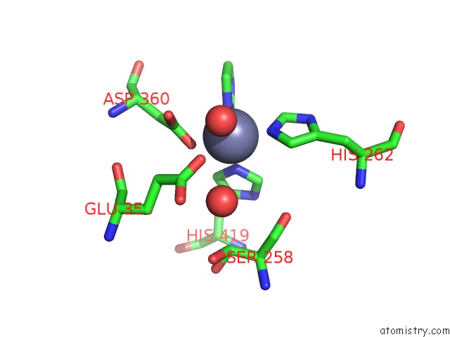
Mono view
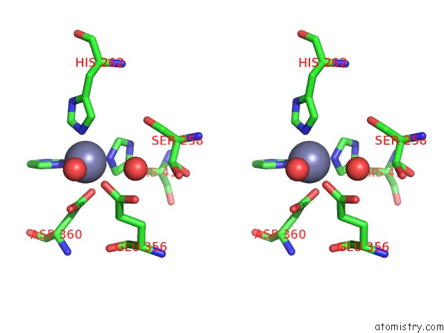
Stereo pair view
Mono view
Stereo pair view
A full contact list of Zinc with other atoms in the Zn binding
site number 2 of L-Histidinol Dehydrogenase (Hisd) Structure Complexed with L- Histidinol (Substrate), Zinc and Nad (Cofactor) within 5.0Å range:
|
Reference:
J.A.R.G.Barbosa,
J.Sivaraman,
Y.Li,
R.Larocque,
A.Matte,
J.D.Schrag,
M.Cygler.
Mechanism of Action and Nad+-Binding Mode Revealed By the Crystal Structure of L-Histidinol Dehydrogenase. Proc.Natl.Acad.Sci.Usa V. 99 1859 2002.
ISSN: ISSN 0027-8424
PubMed: 11842181
DOI: 10.1073/PNAS.022476199
Page generated: Sun Oct 13 04:09:48 2024
ISSN: ISSN 0027-8424
PubMed: 11842181
DOI: 10.1073/PNAS.022476199
Last articles
Mg in 6YARMg in 6YAL
Mg in 6YA8
Mg in 6Y64
Mg in 6Y8D
Mg in 6YA5
Mg in 6Y8E
Mg in 6Y8B
Mg in 6Y8C
Mg in 6Y8A