Zinc »
PDB 1hxy-1i95 »
1i6p »
Zinc in PDB 1i6p: Crystal Structure of E. Coli Beta Carbonic Anhydrase (Ecca)
Enzymatic activity of Crystal Structure of E. Coli Beta Carbonic Anhydrase (Ecca)
All present enzymatic activity of Crystal Structure of E. Coli Beta Carbonic Anhydrase (Ecca):
4.2.1.1;
4.2.1.1;
Protein crystallography data
The structure of Crystal Structure of E. Coli Beta Carbonic Anhydrase (Ecca), PDB code: 1i6p
was solved by
J.D.Cronk,
J.A.Endrizzi,
M.R.Cronk,
J.W.O'neill,
K.Y.J.Zhang,
with X-Ray Crystallography technique. A brief refinement statistics is given in the table below:
| Resolution Low / High (Å) | 42.21 / 2.00 |
| Space group | P 42 21 2 |
| Cell size a, b, c (Å), α, β, γ (°) | 68.535, 68.535, 85.877, 90.00, 90.00, 90.00 |
| R / Rfree (%) | 17.6 / 20.2 |
Zinc Binding Sites:
The binding sites of Zinc atom in the Crystal Structure of E. Coli Beta Carbonic Anhydrase (Ecca)
(pdb code 1i6p). This binding sites where shown within
5.0 Angstroms radius around Zinc atom.
In total only one binding site of Zinc was determined in the Crystal Structure of E. Coli Beta Carbonic Anhydrase (Ecca), PDB code: 1i6p:
In total only one binding site of Zinc was determined in the Crystal Structure of E. Coli Beta Carbonic Anhydrase (Ecca), PDB code: 1i6p:
Zinc binding site 1 out of 1 in 1i6p
Go back to
Zinc binding site 1 out
of 1 in the Crystal Structure of E. Coli Beta Carbonic Anhydrase (Ecca)
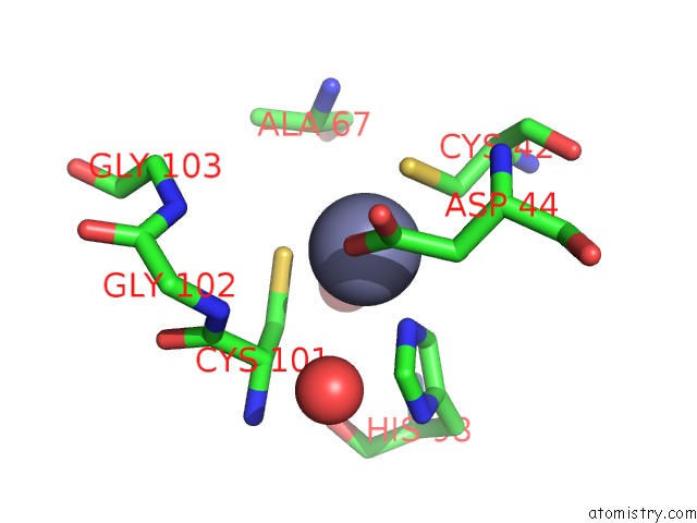
Mono view
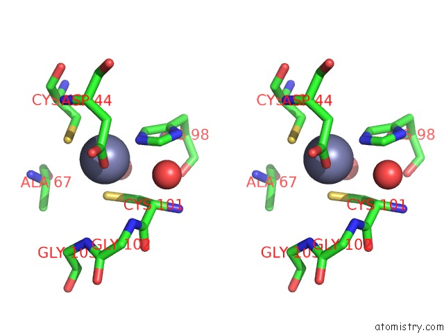
Stereo pair view
Mono view
Stereo pair view
A full contact list of Zinc with other atoms in the Zn binding
site number 1 of Crystal Structure of E. Coli Beta Carbonic Anhydrase (Ecca) within 5.0Å range:
|
Reference:
J.D.Cronk,
J.A.Endrizzi,
M.R.Cronk,
J.W.O'neill,
K.Y.Zhang.
Crystal Structure of E. Coli Beta-Carbonic Anhydrase, An Enzyme with An Unusual pH-Dependent Activity. Protein Sci. V. 10 911 2001.
ISSN: ISSN 0961-8368
PubMed: 11316870
DOI: 10.1110/PS.46301
Page generated: Sun Oct 13 02:53:54 2024
ISSN: ISSN 0961-8368
PubMed: 11316870
DOI: 10.1110/PS.46301
Last articles
Mg in 4PQUMg in 4PQ9
Mg in 4PMW
Mg in 4PPM
Mg in 4PJO
Mg in 4PM0
Mg in 4PL5
Mg in 4PLM
Mg in 4PKO
Mg in 4PKN