Zinc »
PDB 1hlk-1hxr »
1hxp »
Zinc in PDB 1hxp: Nucleotide Transferase
Enzymatic activity of Nucleotide Transferase
All present enzymatic activity of Nucleotide Transferase:
2.7.7.10;
2.7.7.10;
Protein crystallography data
The structure of Nucleotide Transferase, PDB code: 1hxp
was solved by
J.E.Wedekind,
P.A.Frey,
I.Rayment,
with X-Ray Crystallography technique. A brief refinement statistics is given in the table below:
| Resolution Low / High (Å) | 50.00 / 1.80 |
| Space group | P 21 21 2 |
| Cell size a, b, c (Å), α, β, γ (°) | 58.600, 217.200, 69.600, 90.00, 90.00, 90.00 |
| R / Rfree (%) | n/a / n/a |
Other elements in 1hxp:
The structure of Nucleotide Transferase also contains other interesting chemical elements:
| Iron | (Fe) | 2 atoms |
Zinc Binding Sites:
The binding sites of Zinc atom in the Nucleotide Transferase
(pdb code 1hxp). This binding sites where shown within
5.0 Angstroms radius around Zinc atom.
In total 2 binding sites of Zinc where determined in the Nucleotide Transferase, PDB code: 1hxp:
Jump to Zinc binding site number: 1; 2;
In total 2 binding sites of Zinc where determined in the Nucleotide Transferase, PDB code: 1hxp:
Jump to Zinc binding site number: 1; 2;
Zinc binding site 1 out of 2 in 1hxp
Go back to
Zinc binding site 1 out
of 2 in the Nucleotide Transferase
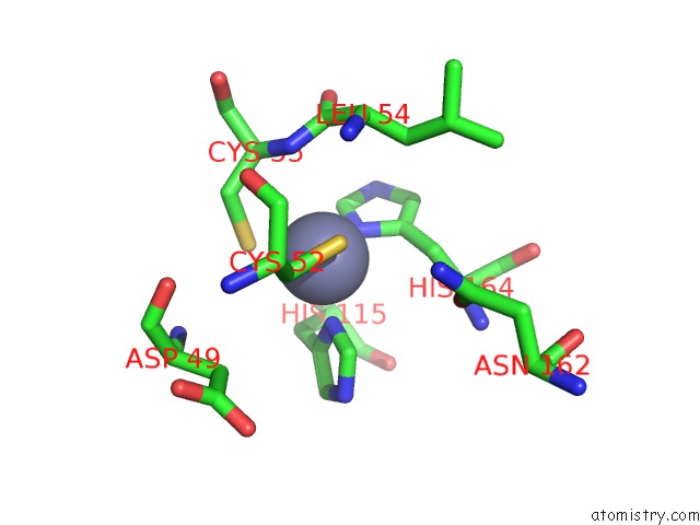
Mono view
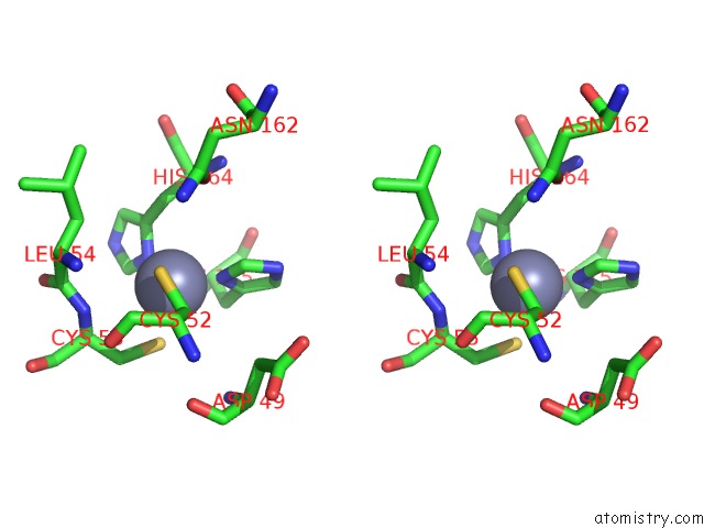
Stereo pair view
Mono view
Stereo pair view
A full contact list of Zinc with other atoms in the Zn binding
site number 1 of Nucleotide Transferase within 5.0Å range:
|
Zinc binding site 2 out of 2 in 1hxp
Go back to
Zinc binding site 2 out
of 2 in the Nucleotide Transferase
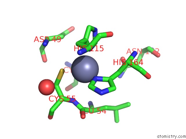
Mono view
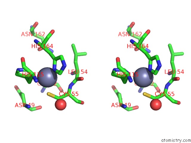
Stereo pair view
Mono view
Stereo pair view
A full contact list of Zinc with other atoms in the Zn binding
site number 2 of Nucleotide Transferase within 5.0Å range:
|
Reference:
J.E.Wedekind,
P.A.Frey,
I.Rayment.
Three-Dimensional Structure of Galactose-1-Phosphate Uridylyltransferase From Escherichia Coli at 1.8 A Resolution. Biochemistry V. 34 11049 1995.
ISSN: ISSN 0006-2960
PubMed: 7669762
DOI: 10.1021/BI00035A010
Page generated: Sun Oct 13 02:37:58 2024
ISSN: ISSN 0006-2960
PubMed: 7669762
DOI: 10.1021/BI00035A010
Last articles
Xe in 1C3LXe in 1C1M
W in 8PRO
W in 9FPP
W in 8PRM
W in 9QM1
W in 9QM0
W in 9OJ3
W in 9MQX
W in 9FP4