Zinc »
PDB 1gkr-1h4t »
1gpc »
Zinc in PDB 1gpc: Core GP32, Dna-Binding Protein
Protein crystallography data
The structure of Core GP32, Dna-Binding Protein, PDB code: 1gpc
was solved by
Y.Shamoo,
A.M.Friedman,
M.R.Parsons,
W.H.Konigsberg,
T.A.Steitz,
with X-Ray Crystallography technique. A brief refinement statistics is given in the table below:
| Resolution Low / High (Å) | 8.00 / 2.20 |
| Space group | P 65 2 2 |
| Cell size a, b, c (Å), α, β, γ (°) | 66.500, 66.500, 235.400, 90.00, 90.00, 120.00 |
| R / Rfree (%) | 23.6 / n/a |
Zinc Binding Sites:
The binding sites of Zinc atom in the Core GP32, Dna-Binding Protein
(pdb code 1gpc). This binding sites where shown within
5.0 Angstroms radius around Zinc atom.
In total only one binding site of Zinc was determined in the Core GP32, Dna-Binding Protein, PDB code: 1gpc:
In total only one binding site of Zinc was determined in the Core GP32, Dna-Binding Protein, PDB code: 1gpc:
Zinc binding site 1 out of 1 in 1gpc
Go back to
Zinc binding site 1 out
of 1 in the Core GP32, Dna-Binding Protein
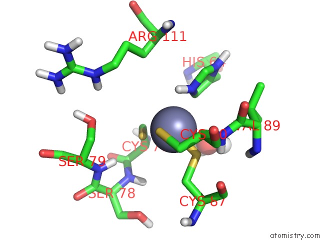
Mono view
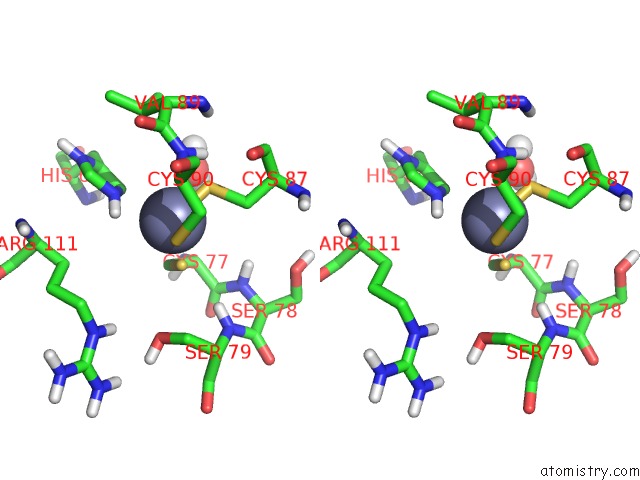
Stereo pair view
Mono view
Stereo pair view
A full contact list of Zinc with other atoms in the Zn binding
site number 1 of Core GP32, Dna-Binding Protein within 5.0Å range:
|
Reference:
Y.Shamoo,
A.M.Friedman,
M.R.Parsons,
W.H.Konigsberg,
T.A.Steitz.
Crystal Structure of A Replication Fork Single-Stranded Dna Binding Protein (T4 GP32) Complexed to Dna. Nature V. 376 362 1995.
ISSN: ISSN 0028-0836
PubMed: 7630406
DOI: 10.1038/376362A0
Page generated: Sun Oct 13 01:39:55 2024
ISSN: ISSN 0028-0836
PubMed: 7630406
DOI: 10.1038/376362A0
Last articles
Na in 6JJENa in 6JIZ
Na in 6JI2
Na in 6JDP
Na in 6JBU
Na in 6JBV
Na in 6JCH
Na in 6JCL
Na in 6J8M
Na in 6JBD