Zinc »
PDB 1g45-1gkq »
1g7a »
Zinc in PDB 1g7a: 1.2 A Structure of T3R3 Human Insulin at 100 K
Protein crystallography data
The structure of 1.2 A Structure of T3R3 Human Insulin at 100 K, PDB code: 1g7a
was solved by
G.D.Smith,
W.A.Pangborn,
R.H.Blessing,
with X-Ray Crystallography technique. A brief refinement statistics is given in the table below:
| Resolution Low / High (Å) | 31.20 / 1.20 |
| Space group | H 3 |
| Cell size a, b, c (Å), α, β, γ (°) | 80.127, 80.127, 71.582, 90.00, 90.00, 120.00 |
| R / Rfree (%) | 16.9 / 19.3 |
Other elements in 1g7a:
The structure of 1.2 A Structure of T3R3 Human Insulin at 100 K also contains other interesting chemical elements:
| Chlorine | (Cl) | 7 atoms |
Zinc Binding Sites:
The binding sites of Zinc atom in the 1.2 A Structure of T3R3 Human Insulin at 100 K
(pdb code 1g7a). This binding sites where shown within
5.0 Angstroms radius around Zinc atom.
In total 7 binding sites of Zinc where determined in the 1.2 A Structure of T3R3 Human Insulin at 100 K, PDB code: 1g7a:
Jump to Zinc binding site number: 1; 2; 3; 4; 5; 6; 7;
In total 7 binding sites of Zinc where determined in the 1.2 A Structure of T3R3 Human Insulin at 100 K, PDB code: 1g7a:
Jump to Zinc binding site number: 1; 2; 3; 4; 5; 6; 7;
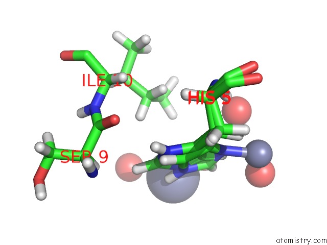
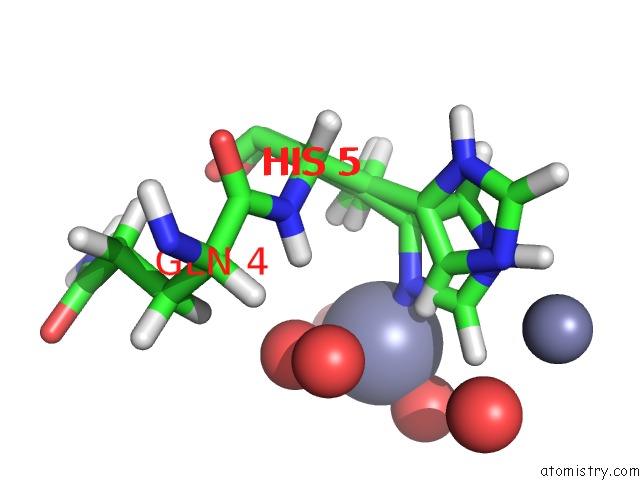
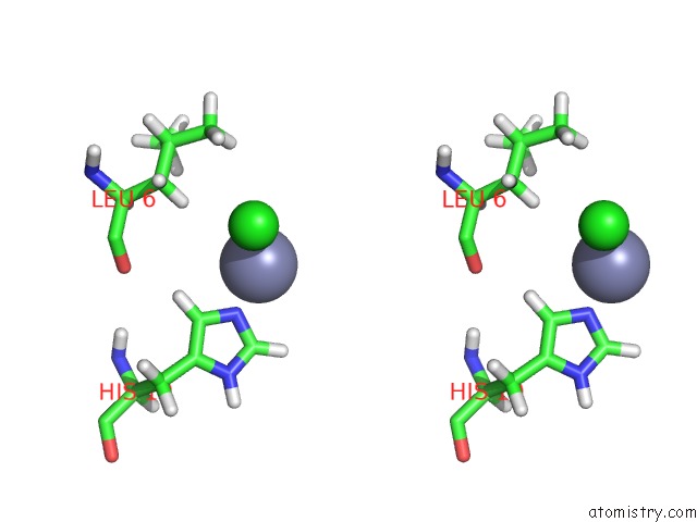
Zinc binding site 1 out of 7 in 1g7a
Go back to
Zinc binding site 1 out
of 7 in the 1.2 A Structure of T3R3 Human Insulin at 100 K

Mono view

Stereo pair view
Mono view
Stereo pair view
A full contact list of Zinc with other atoms in the Zn binding
site number 1 of 1.2 A Structure of T3R3 Human Insulin at 100 K within 5.0Å range:
|
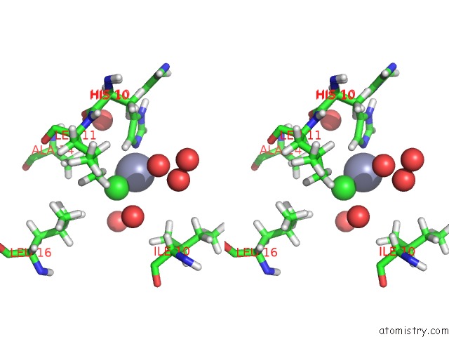
Zinc binding site 2 out of 7 in 1g7a
Go back to
Zinc binding site 2 out
of 7 in the 1.2 A Structure of T3R3 Human Insulin at 100 K
Mono view

Stereo pair view
Mono view
Stereo pair view
A full contact list of Zinc with other atoms in the Zn binding
site number 2 of 1.2 A Structure of T3R3 Human Insulin at 100 K within 5.0Å range:
|
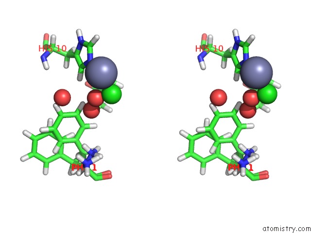
Zinc binding site 3 out of 7 in 1g7a
Go back to
Zinc binding site 3 out
of 7 in the 1.2 A Structure of T3R3 Human Insulin at 100 K
Mono view

Stereo pair view
Mono view
Stereo pair view
A full contact list of Zinc with other atoms in the Zn binding
site number 3 of 1.2 A Structure of T3R3 Human Insulin at 100 K within 5.0Å range:
|
Zinc binding site 4 out of 7 in 1g7a
Go back to
Zinc binding site 4 out
of 7 in the 1.2 A Structure of T3R3 Human Insulin at 100 K

Mono view

Stereo pair view
Mono view
Stereo pair view
A full contact list of Zinc with other atoms in the Zn binding
site number 4 of 1.2 A Structure of T3R3 Human Insulin at 100 K within 5.0Å range:
|
Zinc binding site 5 out of 7 in 1g7a
Go back to
Zinc binding site 5 out
of 7 in the 1.2 A Structure of T3R3 Human Insulin at 100 K

Mono view
Stereo pair view
Mono view
Stereo pair view
A full contact list of Zinc with other atoms in the Zn binding
site number 5 of 1.2 A Structure of T3R3 Human Insulin at 100 K within 5.0Å range:
|
Zinc binding site 6 out of 7 in 1g7a
Go back to
Zinc binding site 6 out
of 7 in the 1.2 A Structure of T3R3 Human Insulin at 100 K

Mono view
Stereo pair view
Mono view
Stereo pair view
A full contact list of Zinc with other atoms in the Zn binding
site number 6 of 1.2 A Structure of T3R3 Human Insulin at 100 K within 5.0Å range:
|
Zinc binding site 7 out of 7 in 1g7a
Go back to
Zinc binding site 7 out
of 7 in the 1.2 A Structure of T3R3 Human Insulin at 100 K

Mono view
Stereo pair view
Mono view
Stereo pair view
A full contact list of Zinc with other atoms in the Zn binding
site number 7 of 1.2 A Structure of T3R3 Human Insulin at 100 K within 5.0Å range:
|
Reference:
G.D.Smith,
W.A.Pangborn,
R.H.Blessing.
Phase Changes in T(3)R(3)(F) Human Insulin: Temperature or Pressure Induced? Acta Crystallogr.,Sect.D V. 57 1091 2001.
ISSN: ISSN 0907-4449
PubMed: 11468392
DOI: 10.1107/S0907444901007685
Page generated: Sun Oct 13 01:23:30 2024
ISSN: ISSN 0907-4449
PubMed: 11468392
DOI: 10.1107/S0907444901007685
Last articles
Fe in 6ZKLFe in 6ZKI
Fe in 6ZKK
Fe in 6ZKJ
Fe in 6ZKH
Fe in 6ZKG
Fe in 6ZKF
Fe in 6ZKE
Fe in 6ZKD
Fe in 6ZKC