Zinc »
PDB 1g45-1gkq »
1g71 »
Zinc in PDB 1g71: Crystal Structure of Pyrococcus Furiosus Dna Primase
Protein crystallography data
The structure of Crystal Structure of Pyrococcus Furiosus Dna Primase, PDB code: 1g71
was solved by
M.A.Augustin,
R.Huber,
J.T.Kaiser,
with X-Ray Crystallography technique. A brief refinement statistics is given in the table below:
| Resolution Low / High (Å) | 500.00 / 2.30 |
| Space group | P 21 21 2 |
| Cell size a, b, c (Å), α, β, γ (°) | 129.883, 136.326, 61.877, 90.00, 90.00, 90.00 |
| R / Rfree (%) | 21.1 / 24.9 |
Other elements in 1g71:
The structure of Crystal Structure of Pyrococcus Furiosus Dna Primase also contains other interesting chemical elements:
| Chlorine | (Cl) | 6 atoms |
Zinc Binding Sites:
The binding sites of Zinc atom in the Crystal Structure of Pyrococcus Furiosus Dna Primase
(pdb code 1g71). This binding sites where shown within
5.0 Angstroms radius around Zinc atom.
In total 2 binding sites of Zinc where determined in the Crystal Structure of Pyrococcus Furiosus Dna Primase, PDB code: 1g71:
Jump to Zinc binding site number: 1; 2;
In total 2 binding sites of Zinc where determined in the Crystal Structure of Pyrococcus Furiosus Dna Primase, PDB code: 1g71:
Jump to Zinc binding site number: 1; 2;
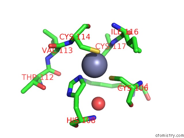
Zinc binding site 1 out of 2 in 1g71
Go back to
Zinc binding site 1 out
of 2 in the Crystal Structure of Pyrococcus Furiosus Dna Primase
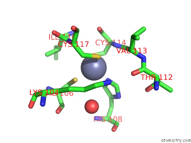
Mono view
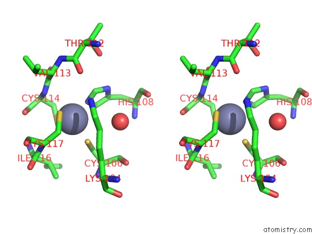
Stereo pair view
Mono view
Stereo pair view
A full contact list of Zinc with other atoms in the Zn binding
site number 1 of Crystal Structure of Pyrococcus Furiosus Dna Primase within 5.0Å range:
|
Zinc binding site 2 out of 2 in 1g71
Go back to
Zinc binding site 2 out
of 2 in the Crystal Structure of Pyrococcus Furiosus Dna Primase
Mono view

Stereo pair view
Mono view
Stereo pair view
A full contact list of Zinc with other atoms in the Zn binding
site number 2 of Crystal Structure of Pyrococcus Furiosus Dna Primase within 5.0Å range:
|
Reference:
M.A.Augustin,
R.Huber,
J.T.Kaiser.
Crystal Structure of A Dna-Dependent Rna Polymerase (Dna Primase). Nat.Struct.Biol. V. 8 57 2001.
ISSN: ISSN 1072-8368
PubMed: 11135672
DOI: 10.1038/83060
Page generated: Sun Oct 13 01:22:55 2024
ISSN: ISSN 1072-8368
PubMed: 11135672
DOI: 10.1038/83060
Last articles
Fe in 6ZKPFe in 6ZKO
Fe in 6ZKM
Fe in 6ZKN
Fe in 6ZKL
Fe in 6ZKI
Fe in 6ZKK
Fe in 6ZKJ
Fe in 6ZKH
Fe in 6ZKG