Zinc »
PDB 1fpp-1g43 »
1fss »
Zinc in PDB 1fss: Acetylcholinesterase (E.C. 3.1.1.7) Complexed with Fasciculin-II
Enzymatic activity of Acetylcholinesterase (E.C. 3.1.1.7) Complexed with Fasciculin-II
All present enzymatic activity of Acetylcholinesterase (E.C. 3.1.1.7) Complexed with Fasciculin-II:
3.1.1.7;
3.1.1.7;
Protein crystallography data
The structure of Acetylcholinesterase (E.C. 3.1.1.7) Complexed with Fasciculin-II, PDB code: 1fss
was solved by
M.Harel,
G.J.Kleywegt,
I.Silman,
J.L.Sussman,
with X-Ray Crystallography technique. A brief refinement statistics is given in the table below:
| Resolution Low / High (Å) | 8.00 / 3.00 |
| Space group | P 21 21 2 |
| Cell size a, b, c (Å), α, β, γ (°) | 87.390, 115.000, 67.470, 90.00, 90.00, 90.00 |
| R / Rfree (%) | 23 / 31 |
Zinc Binding Sites:
The binding sites of Zinc atom in the Acetylcholinesterase (E.C. 3.1.1.7) Complexed with Fasciculin-II
(pdb code 1fss). This binding sites where shown within
5.0 Angstroms radius around Zinc atom.
In total 2 binding sites of Zinc where determined in the Acetylcholinesterase (E.C. 3.1.1.7) Complexed with Fasciculin-II, PDB code: 1fss:
Jump to Zinc binding site number: 1; 2;
In total 2 binding sites of Zinc where determined in the Acetylcholinesterase (E.C. 3.1.1.7) Complexed with Fasciculin-II, PDB code: 1fss:
Jump to Zinc binding site number: 1; 2;
Zinc binding site 1 out of 2 in 1fss
Go back to
Zinc binding site 1 out
of 2 in the Acetylcholinesterase (E.C. 3.1.1.7) Complexed with Fasciculin-II
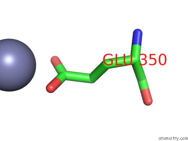
Mono view
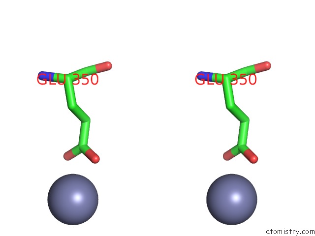
Stereo pair view
Mono view
Stereo pair view
A full contact list of Zinc with other atoms in the Zn binding
site number 1 of Acetylcholinesterase (E.C. 3.1.1.7) Complexed with Fasciculin-II within 5.0Å range:
|
Zinc binding site 2 out of 2 in 1fss
Go back to
Zinc binding site 2 out
of 2 in the Acetylcholinesterase (E.C. 3.1.1.7) Complexed with Fasciculin-II
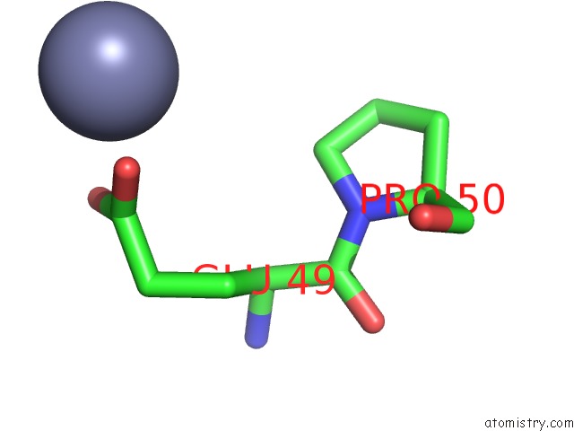
Mono view
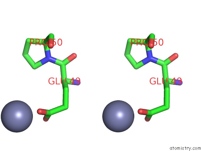
Stereo pair view
Mono view
Stereo pair view
A full contact list of Zinc with other atoms in the Zn binding
site number 2 of Acetylcholinesterase (E.C. 3.1.1.7) Complexed with Fasciculin-II within 5.0Å range:
|
Reference:
M.Harel,
G.J.Kleywegt,
R.B.Ravelli,
I.Silman,
J.L.Sussman.
Crystal Structure of An Acetylcholinesterase-Fasciculin Complex: Interaction of A Three-Fingered Toxin From Snake Venom with Its Target. Structure V. 3 1355 1995.
ISSN: ISSN 0969-2126
PubMed: 8747462
DOI: 10.1016/S0969-2126(01)00273-8
Page generated: Sun Oct 13 01:07:13 2024
ISSN: ISSN 0969-2126
PubMed: 8747462
DOI: 10.1016/S0969-2126(01)00273-8
Last articles
Mn in 5RAAMn in 5OMX
Mn in 5OXJ
Mn in 5R7X
Mn in 5OX6
Mn in 5OX5
Mn in 5OR6
Mn in 5OVO
Mn in 5OR2
Mn in 5ONW