Zinc »
PDB 1f6u-1fp0 »
1fno »
Zinc in PDB 1fno: Peptidase T (Tripeptidase)
Protein crystallography data
The structure of Peptidase T (Tripeptidase), PDB code: 1fno
was solved by
K.Hakansson,
C.G.Miller,
with X-Ray Crystallography technique. A brief refinement statistics is given in the table below:
| Resolution Low / High (Å) | 500.00 / 2.40 |
| Space group | C 1 2 1 |
| Cell size a, b, c (Å), α, β, γ (°) | 132.374, 46.036, 96.591, 90.00, 116.11, 90.00 |
| R / Rfree (%) | 22.2 / 26.1 |
Zinc Binding Sites:
The binding sites of Zinc atom in the Peptidase T (Tripeptidase)
(pdb code 1fno). This binding sites where shown within
5.0 Angstroms radius around Zinc atom.
In total 2 binding sites of Zinc where determined in the Peptidase T (Tripeptidase), PDB code: 1fno:
Jump to Zinc binding site number: 1; 2;
In total 2 binding sites of Zinc where determined in the Peptidase T (Tripeptidase), PDB code: 1fno:
Jump to Zinc binding site number: 1; 2;
Zinc binding site 1 out of 2 in 1fno
Go back to
Zinc binding site 1 out
of 2 in the Peptidase T (Tripeptidase)
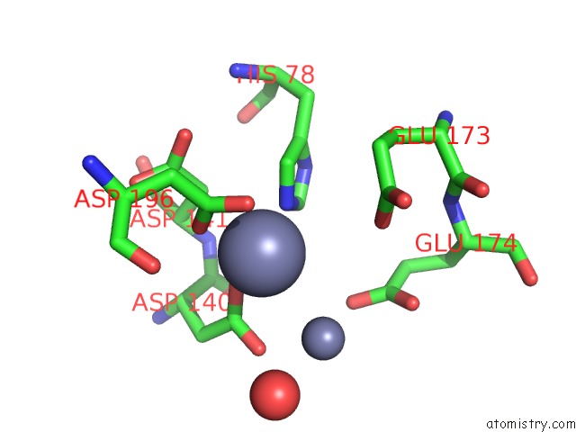
Mono view
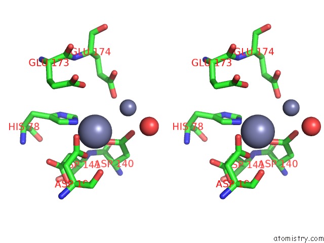
Stereo pair view
Mono view
Stereo pair view
A full contact list of Zinc with other atoms in the Zn binding
site number 1 of Peptidase T (Tripeptidase) within 5.0Å range:
|
Zinc binding site 2 out of 2 in 1fno
Go back to
Zinc binding site 2 out
of 2 in the Peptidase T (Tripeptidase)
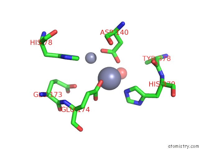
Mono view
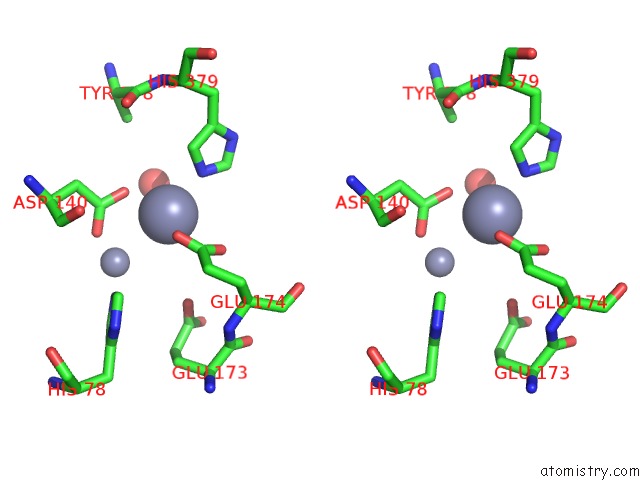
Stereo pair view
Mono view
Stereo pair view
A full contact list of Zinc with other atoms in the Zn binding
site number 2 of Peptidase T (Tripeptidase) within 5.0Å range:
|
Reference:
K.Hakansson,
C.G.Miller.
Structure of Peptidase T From Salmonella Typhimurium Eur.J.Biochem. V. 269 443 2002.
ISSN: ISSN 0014-2956
PubMed: 11856302
DOI: 10.1046/J.0014-2956.2001.02665.X
Page generated: Sun Oct 13 01:02:53 2024
ISSN: ISSN 0014-2956
PubMed: 11856302
DOI: 10.1046/J.0014-2956.2001.02665.X
Last articles
Mg in 5T41Mg in 5T45
Mg in 5T3K
Mg in 5T14
Mg in 5SZT
Mg in 5T2V
Mg in 5T13
Mg in 5T19
Mg in 5SZK
Mg in 5SZJ