Zinc »
PDB 1evr-1f62 »
1ezz »
Zinc in PDB 1ezz: Crystal Structure of E. Coli Aspartate Transcarbamoylase P268A Mutant in the T-State
Enzymatic activity of Crystal Structure of E. Coli Aspartate Transcarbamoylase P268A Mutant in the T-State
All present enzymatic activity of Crystal Structure of E. Coli Aspartate Transcarbamoylase P268A Mutant in the T-State:
2.1.3.2;
2.1.3.2;
Protein crystallography data
The structure of Crystal Structure of E. Coli Aspartate Transcarbamoylase P268A Mutant in the T-State, PDB code: 1ezz
was solved by
L.Jin,
B.Stec,
E.R.Kantrowitz,
with X-Ray Crystallography technique. A brief refinement statistics is given in the table below:
| Resolution Low / High (Å) | 8.00 / 2.70 |
| Space group | H 3 |
| Cell size a, b, c (Å), α, β, γ (°) | 129.870, 129.870, 198.340, 90.00, 90.00, 120.00 |
| R / Rfree (%) | 18.2 / 24.2 |
Zinc Binding Sites:
The binding sites of Zinc atom in the Crystal Structure of E. Coli Aspartate Transcarbamoylase P268A Mutant in the T-State
(pdb code 1ezz). This binding sites where shown within
5.0 Angstroms radius around Zinc atom.
In total 2 binding sites of Zinc where determined in the Crystal Structure of E. Coli Aspartate Transcarbamoylase P268A Mutant in the T-State, PDB code: 1ezz:
Jump to Zinc binding site number: 1; 2;
In total 2 binding sites of Zinc where determined in the Crystal Structure of E. Coli Aspartate Transcarbamoylase P268A Mutant in the T-State, PDB code: 1ezz:
Jump to Zinc binding site number: 1; 2;
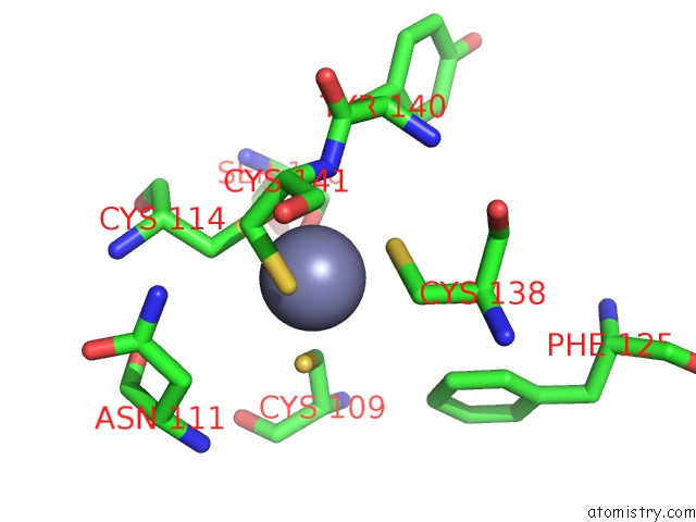
Zinc binding site 1 out of 2 in 1ezz
Go back to
Zinc binding site 1 out
of 2 in the Crystal Structure of E. Coli Aspartate Transcarbamoylase P268A Mutant in the T-State

Mono view

Stereo pair view
Mono view
Stereo pair view
A full contact list of Zinc with other atoms in the Zn binding
site number 1 of Crystal Structure of E. Coli Aspartate Transcarbamoylase P268A Mutant in the T-State within 5.0Å range:
|
Zinc binding site 2 out of 2 in 1ezz
Go back to
Zinc binding site 2 out
of 2 in the Crystal Structure of E. Coli Aspartate Transcarbamoylase P268A Mutant in the T-State
Mono view

Stereo pair view
Mono view
Stereo pair view
A full contact list of Zinc with other atoms in the Zn binding
site number 2 of Crystal Structure of E. Coli Aspartate Transcarbamoylase P268A Mutant in the T-State within 5.0Å range:
|
Reference:
L.Jin,
B.Stec,
E.R.Kantrowitz.
A Cis-Proline to Alanine Mutant of E. Coli Aspartate Transcarbamoylase: Kinetic Studies and Three-Dimensional Crystal Structures. Biochemistry V. 39 8058 2000.
ISSN: ISSN 0006-2960
PubMed: 10891088
DOI: 10.1021/BI000418+
Page generated: Sun Oct 13 00:36:03 2024
ISSN: ISSN 0006-2960
PubMed: 10891088
DOI: 10.1021/BI000418+
Last articles
Na in 6JJENa in 6JIZ
Na in 6JI2
Na in 6JDP
Na in 6JBU
Na in 6JBV
Na in 6JCH
Na in 6JCL
Na in 6J8M
Na in 6JBD