Zinc »
PDB 1e0e-1ed6 »
1eb0 »
Zinc in PDB 1eb0: Crystal Structure of Bacillus Pasteurii Uree at 1.85 A, Phased By Siras. Type I Crystal Form.
Protein crystallography data
The structure of Crystal Structure of Bacillus Pasteurii Uree at 1.85 A, Phased By Siras. Type I Crystal Form., PDB code: 1eb0
was solved by
H.Remaut,
N.Safarov,
S.Ciurli,
J.Van Beeumen,
with X-Ray Crystallography technique. A brief refinement statistics is given in the table below:
| Resolution Low / High (Å) | 14.85 / 1.85 |
| Space group | I 2 2 2 |
| Cell size a, b, c (Å), α, β, γ (°) | 50.500, 60.720, 130.580, 90.00, 90.00, 90.00 |
| R / Rfree (%) | 22 / 25.6 |
Zinc Binding Sites:
The binding sites of Zinc atom in the Crystal Structure of Bacillus Pasteurii Uree at 1.85 A, Phased By Siras. Type I Crystal Form.
(pdb code 1eb0). This binding sites where shown within
5.0 Angstroms radius around Zinc atom.
In total only one binding site of Zinc was determined in the Crystal Structure of Bacillus Pasteurii Uree at 1.85 A, Phased By Siras. Type I Crystal Form., PDB code: 1eb0:
In total only one binding site of Zinc was determined in the Crystal Structure of Bacillus Pasteurii Uree at 1.85 A, Phased By Siras. Type I Crystal Form., PDB code: 1eb0:
Zinc binding site 1 out of 1 in 1eb0
Go back to
Zinc binding site 1 out
of 1 in the Crystal Structure of Bacillus Pasteurii Uree at 1.85 A, Phased By Siras. Type I Crystal Form.
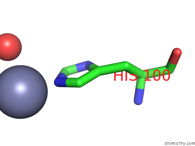
Mono view
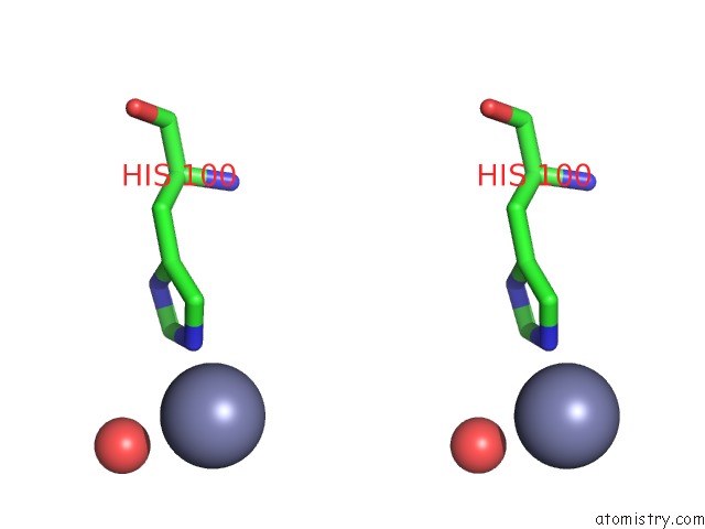
Stereo pair view
Mono view
Stereo pair view
A full contact list of Zinc with other atoms in the Zn binding
site number 1 of Crystal Structure of Bacillus Pasteurii Uree at 1.85 A, Phased By Siras. Type I Crystal Form. within 5.0Å range:
|
Reference:
H.Reamut,
N.Safarov,
S.Ciurli,
J.Van Beeumen.
Structural Basis For NI2+ Transport and Assembly of the Urease Active Site By the Metallochaperone Uree From Bacillus Pasteurii J.Biol.Chem. V. 276 49365 2001.
ISSN: ISSN 0021-9258
PubMed: 11602602
DOI: 10.1074/JBC.M108304200
Page generated: Sun Oct 13 00:06:36 2024
ISSN: ISSN 0021-9258
PubMed: 11602602
DOI: 10.1074/JBC.M108304200
Last articles
Mg in 4R18Mg in 4R17
Mg in 4R4P
Mg in 4R47
Mg in 4R39
Mg in 4R3A
Mg in 4R1F
Mg in 4R2M
Mg in 4R2L
Mg in 4R0D