Zinc »
PDB 1e0e-1ed6 »
1e51 »
Zinc in PDB 1e51: Crystal Structure of Native Human Erythrocyte 5-Aminolaevulinic Acid Dehydratase
Enzymatic activity of Crystal Structure of Native Human Erythrocyte 5-Aminolaevulinic Acid Dehydratase
All present enzymatic activity of Crystal Structure of Native Human Erythrocyte 5-Aminolaevulinic Acid Dehydratase:
4.2.1.24;
4.2.1.24;
Protein crystallography data
The structure of Crystal Structure of Native Human Erythrocyte 5-Aminolaevulinic Acid Dehydratase, PDB code: 1e51
was solved by
N.L.Mills-Davies,
D.Thompson,
J.B.Cooper,
P.M.Shoolingin-Jordan,
with X-Ray Crystallography technique. A brief refinement statistics is given in the table below:
| Resolution Low / High (Å) | 44.38 / 2.83 |
| Space group | I 4 2 2 |
| Cell size a, b, c (Å), α, β, γ (°) | 125.530, 125.530, 200.910, 90.00, 90.00, 90.00 |
| R / Rfree (%) | 21.3 / 27 |
Zinc Binding Sites:
The binding sites of Zinc atom in the Crystal Structure of Native Human Erythrocyte 5-Aminolaevulinic Acid Dehydratase
(pdb code 1e51). This binding sites where shown within
5.0 Angstroms radius around Zinc atom.
In total only one binding site of Zinc was determined in the Crystal Structure of Native Human Erythrocyte 5-Aminolaevulinic Acid Dehydratase, PDB code: 1e51:
In total only one binding site of Zinc was determined in the Crystal Structure of Native Human Erythrocyte 5-Aminolaevulinic Acid Dehydratase, PDB code: 1e51:
Zinc binding site 1 out of 1 in 1e51
Go back to
Zinc binding site 1 out
of 1 in the Crystal Structure of Native Human Erythrocyte 5-Aminolaevulinic Acid Dehydratase

Mono view
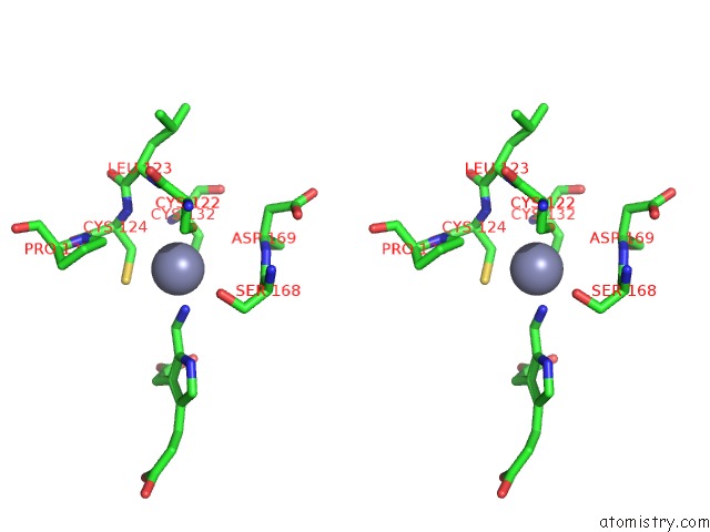
Stereo pair view
Mono view
Stereo pair view
A full contact list of Zinc with other atoms in the Zn binding
site number 1 of Crystal Structure of Native Human Erythrocyte 5-Aminolaevulinic Acid Dehydratase within 5.0Å range:
|
Reference:
N.L.Mills-Davies,
D.Thompson,
J.B.Cooper,
S.P.Wood,
P.M.Shoolingin-Jordan.
The Crystal Structure of Human Ala-Dehydratase To Be Published.
Page generated: Sun Oct 13 00:03:16 2024
Last articles
Mg in 3CUNMg in 3CUS
Mg in 3CUL
Mg in 3CUR
Mg in 3CTL
Mg in 3CT7
Mg in 3CU8
Mg in 3CU3
Mg in 3CT2
Mg in 3CSK