Zinc »
PDB 1caq-1co4 »
1clc »
Zinc in PDB 1clc: Three-Dimensional Structure of Endoglucanase D at 1.9 Angstroms Resolution
Enzymatic activity of Three-Dimensional Structure of Endoglucanase D at 1.9 Angstroms Resolution
All present enzymatic activity of Three-Dimensional Structure of Endoglucanase D at 1.9 Angstroms Resolution:
3.2.1.4;
3.2.1.4;
Protein crystallography data
The structure of Three-Dimensional Structure of Endoglucanase D at 1.9 Angstroms Resolution, PDB code: 1clc
was solved by
P.M.Alzari,
M.B.Lascombe,
with X-Ray Crystallography technique. A brief refinement statistics is given in the table below:
| Resolution Low / High (Å) | 7.00 / 1.90 |
| Space group | P 31 2 1 |
| Cell size a, b, c (Å), α, β, γ (°) | 98.900, 98.900, 191.400, 90.00, 90.00, 120.00 |
| R / Rfree (%) | 20.4 / n/a |
Other elements in 1clc:
The structure of Three-Dimensional Structure of Endoglucanase D at 1.9 Angstroms Resolution also contains other interesting chemical elements:
| Calcium | (Ca) | 3 atoms |
Zinc Binding Sites:
The binding sites of Zinc atom in the Three-Dimensional Structure of Endoglucanase D at 1.9 Angstroms Resolution
(pdb code 1clc). This binding sites where shown within
5.0 Angstroms radius around Zinc atom.
In total only one binding site of Zinc was determined in the Three-Dimensional Structure of Endoglucanase D at 1.9 Angstroms Resolution, PDB code: 1clc:
In total only one binding site of Zinc was determined in the Three-Dimensional Structure of Endoglucanase D at 1.9 Angstroms Resolution, PDB code: 1clc:
Zinc binding site 1 out of 1 in 1clc
Go back to
Zinc binding site 1 out
of 1 in the Three-Dimensional Structure of Endoglucanase D at 1.9 Angstroms Resolution
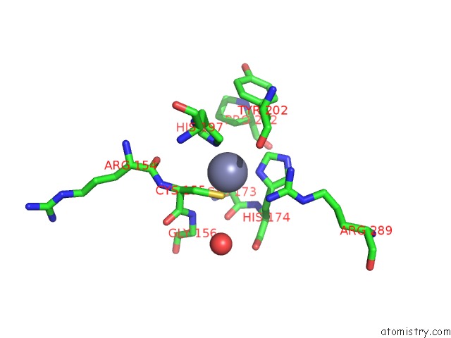
Mono view
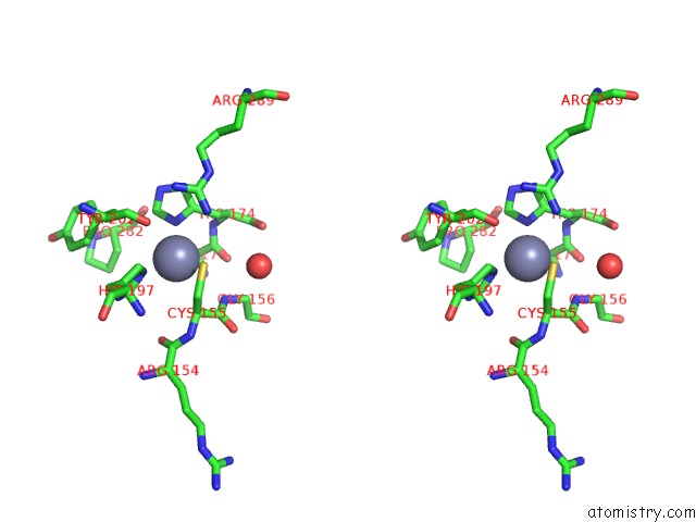
Stereo pair view
Mono view
Stereo pair view
A full contact list of Zinc with other atoms in the Zn binding
site number 1 of Three-Dimensional Structure of Endoglucanase D at 1.9 Angstroms Resolution within 5.0Å range:
|
Reference:
M.B.Lascombe,
H.Souchon,
M.Juy,
P.M.Alzari.
Three-Dimensional Structure of Endoglucanase D at 1.9 Angstroms Resolution To Be Published.
Page generated: Sat Oct 12 23:12:08 2024
Last articles
Ni in 5D1RNi in 5D3P
Ni in 5D27
Ni in 5CK0
Ni in 5CLK
Ni in 5CXD
Ni in 5CK6
Ni in 5C74
Ni in 5C3S
Ni in 5CEH