Zinc »
PDB 1bj6-1bv3 »
1bm6 »
Zinc in PDB 1bm6: Solution Structure of the Catalytic Domain of Human Stromelysin-1 Complexed to A Potent Non-Peptidic Inhibitor, uc(Nmr), 20 Structures
Enzymatic activity of Solution Structure of the Catalytic Domain of Human Stromelysin-1 Complexed to A Potent Non-Peptidic Inhibitor, uc(Nmr), 20 Structures
All present enzymatic activity of Solution Structure of the Catalytic Domain of Human Stromelysin-1 Complexed to A Potent Non-Peptidic Inhibitor, uc(Nmr), 20 Structures:
3.4.24.17;
3.4.24.17;
Other elements in 1bm6:
The structure of Solution Structure of the Catalytic Domain of Human Stromelysin-1 Complexed to A Potent Non-Peptidic Inhibitor, uc(Nmr), 20 Structures also contains other interesting chemical elements:
| Calcium | (Ca) | 40 atoms |
Zinc Binding Sites:
The binding sites of Zinc atom in the Solution Structure of the Catalytic Domain of Human Stromelysin-1 Complexed to A Potent Non-Peptidic Inhibitor, uc(Nmr), 20 Structures
(pdb code 1bm6). This binding sites where shown within
5.0 Angstroms radius around Zinc atom.
In total 2 binding sites of Zinc where determined in the Solution Structure of the Catalytic Domain of Human Stromelysin-1 Complexed to A Potent Non-Peptidic Inhibitor, uc(Nmr), 20 Structures, PDB code: 1bm6:
Jump to Zinc binding site number: 1; 2;
In total 2 binding sites of Zinc where determined in the Solution Structure of the Catalytic Domain of Human Stromelysin-1 Complexed to A Potent Non-Peptidic Inhibitor, uc(Nmr), 20 Structures, PDB code: 1bm6:
Jump to Zinc binding site number: 1; 2;
Zinc binding site 1 out of 2 in 1bm6
Go back to
Zinc binding site 1 out
of 2 in the Solution Structure of the Catalytic Domain of Human Stromelysin-1 Complexed to A Potent Non-Peptidic Inhibitor, uc(Nmr), 20 Structures
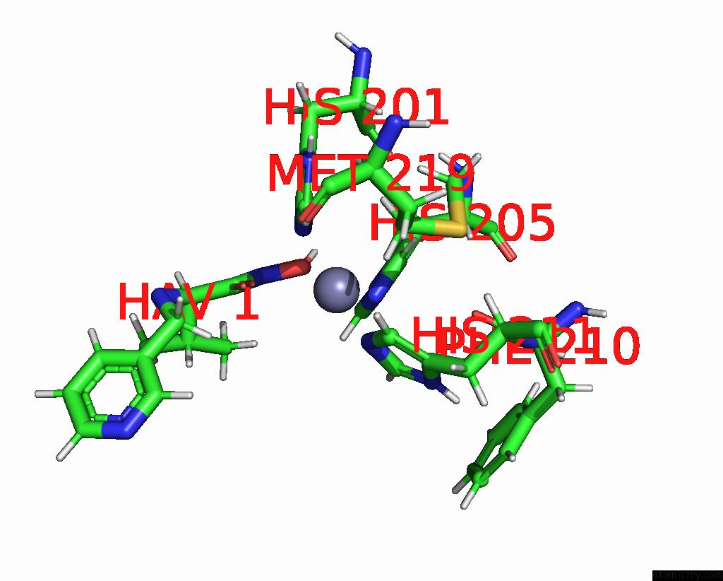
Mono view
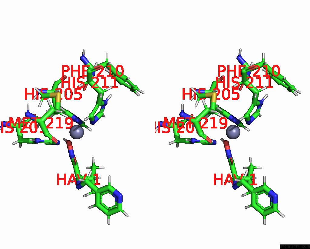
Stereo pair view
Mono view
Stereo pair view
A full contact list of Zinc with other atoms in the Zn binding
site number 1 of Solution Structure of the Catalytic Domain of Human Stromelysin-1 Complexed to A Potent Non-Peptidic Inhibitor, uc(Nmr), 20 Structures within 5.0Å range:
|
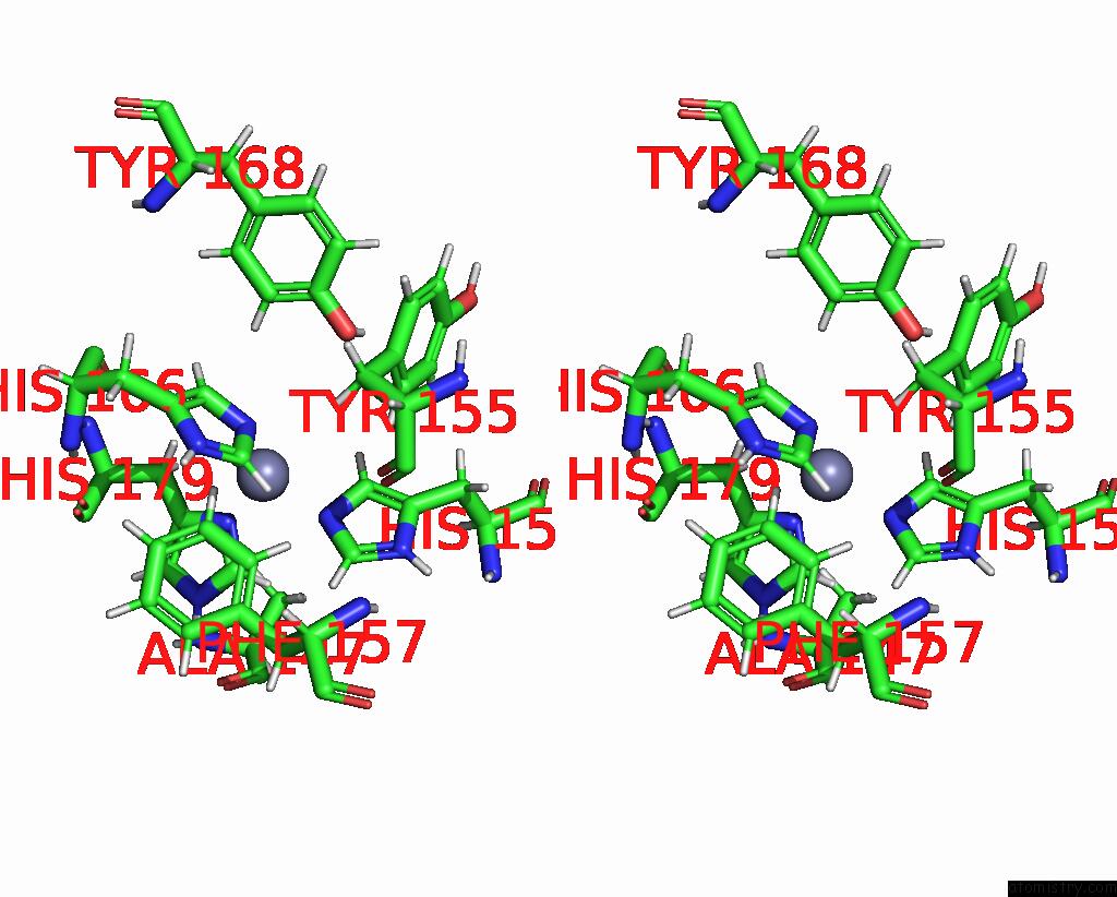
Zinc binding site 2 out of 2 in 1bm6
Go back to
Zinc binding site 2 out
of 2 in the Solution Structure of the Catalytic Domain of Human Stromelysin-1 Complexed to A Potent Non-Peptidic Inhibitor, uc(Nmr), 20 Structures

Mono view
Stereo pair view
Mono view
Stereo pair view
A full contact list of Zinc with other atoms in the Zn binding
site number 2 of Solution Structure of the Catalytic Domain of Human Stromelysin-1 Complexed to A Potent Non-Peptidic Inhibitor, uc(Nmr), 20 Structures within 5.0Å range:
|
Reference:
Y.C.Li,
X.Zhang,
R.Melton,
V.Ganu,
N.C.Gonnella.
Solution Structure of the Catalytic Domain of Human Stromelysin-1 Complexed to A Potent, Nonpeptidic Inhibitor. Biochemistry V. 37 14048 1998.
ISSN: ISSN 0006-2960
PubMed: 9760240
DOI: 10.1021/BI981328W
Page generated: Sat Oct 12 22:35:23 2024
ISSN: ISSN 0006-2960
PubMed: 9760240
DOI: 10.1021/BI981328W
Last articles
Mg in 6YBWMg in 6YKW
Mg in 6YKV
Mg in 6YKU
Mg in 6YKT
Mg in 6YKS
Mg in 6YKQ
Mg in 6YKO
Mg in 6YKN
Mg in 6YKL