Zinc »
PDB 1adf-1axg »
1anv »
Zinc in PDB 1anv: Adenovirus 5 Dbp/Uranyl Fluoride Soak
Protein crystallography data
The structure of Adenovirus 5 Dbp/Uranyl Fluoride Soak, PDB code: 1anv
was solved by
P.N.Kanellopoulos,
D.Tsernoglou,
P.C.Van Der Vliet,
P.A.Tucker,
with X-Ray Crystallography technique. A brief refinement statistics is given in the table below:
| Resolution Low / High (Å) | 15.00 / 2.70 |
| Space group | P 21 21 21 |
| Cell size a, b, c (Å), α, β, γ (°) | 79.700, 75.600, 60.600, 90.00, 90.00, 90.00 |
| R / Rfree (%) | 20.4 / 31.4 |
Other elements in 1anv:
The structure of Adenovirus 5 Dbp/Uranyl Fluoride Soak also contains other interesting chemical elements:
| Uranium | (U) | 4 atoms |
Zinc Binding Sites:
The binding sites of Zinc atom in the Adenovirus 5 Dbp/Uranyl Fluoride Soak
(pdb code 1anv). This binding sites where shown within
5.0 Angstroms radius around Zinc atom.
In total 2 binding sites of Zinc where determined in the Adenovirus 5 Dbp/Uranyl Fluoride Soak, PDB code: 1anv:
Jump to Zinc binding site number: 1; 2;
In total 2 binding sites of Zinc where determined in the Adenovirus 5 Dbp/Uranyl Fluoride Soak, PDB code: 1anv:
Jump to Zinc binding site number: 1; 2;
Zinc binding site 1 out of 2 in 1anv
Go back to
Zinc binding site 1 out
of 2 in the Adenovirus 5 Dbp/Uranyl Fluoride Soak
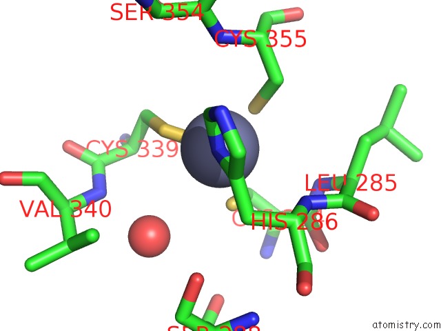
Mono view
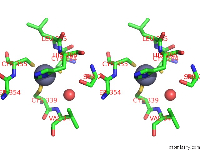
Stereo pair view
Mono view
Stereo pair view
A full contact list of Zinc with other atoms in the Zn binding
site number 1 of Adenovirus 5 Dbp/Uranyl Fluoride Soak within 5.0Å range:
|
Zinc binding site 2 out of 2 in 1anv
Go back to
Zinc binding site 2 out
of 2 in the Adenovirus 5 Dbp/Uranyl Fluoride Soak
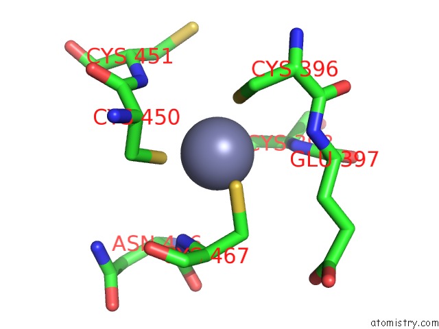
Mono view
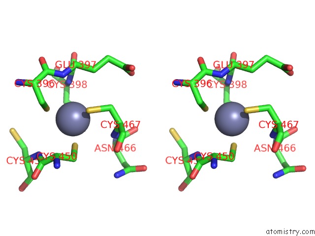
Stereo pair view
Mono view
Stereo pair view
A full contact list of Zinc with other atoms in the Zn binding
site number 2 of Adenovirus 5 Dbp/Uranyl Fluoride Soak within 5.0Å range:
|
Reference:
P.N.Kanellopoulos,
D.Tsernoglou,
P.C.Van Der Vliet,
P.A.Tucker.
Conformational Change of the Adenovirus Dna-Binding Protein Induced By Soaking Crystals with K3UO2F5 Solutions. Acta Crystallogr.,Sect.D V. 52 942 1996.
ISSN: ISSN 0907-4449
PubMed: 15299602
DOI: 10.1107/S0907444996005525
Page generated: Tue Aug 19 19:12:37 2025
ISSN: ISSN 0907-4449
PubMed: 15299602
DOI: 10.1107/S0907444996005525
Last articles
Mn in 9LJUMn in 9LJW
Mn in 9LJS
Mn in 9LJR
Mn in 9LJT
Mn in 9LJV
Mg in 9UA2
Mg in 9R96
Mg in 9VM1
Mg in 9P01