Zinc »
PDB 1adf-1axg »
1aiy »
Zinc in PDB 1aiy: R6 Human Insulin Hexamer (Symmetric), uc(Nmr), 10 Structures
Zinc Binding Sites:
The binding sites of Zinc atom in the R6 Human Insulin Hexamer (Symmetric), uc(Nmr), 10 Structures
(pdb code 1aiy). This binding sites where shown within
5.0 Angstroms radius around Zinc atom.
In total 2 binding sites of Zinc where determined in the R6 Human Insulin Hexamer (Symmetric), uc(Nmr), 10 Structures, PDB code: 1aiy:
Jump to Zinc binding site number: 1; 2;
In total 2 binding sites of Zinc where determined in the R6 Human Insulin Hexamer (Symmetric), uc(Nmr), 10 Structures, PDB code: 1aiy:
Jump to Zinc binding site number: 1; 2;
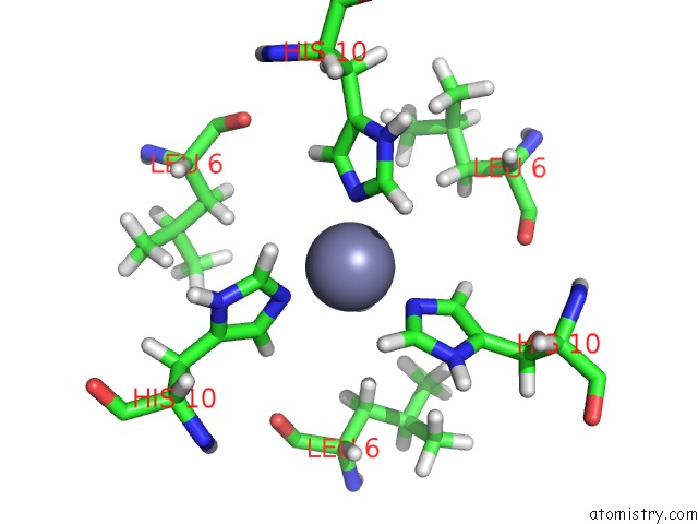
Zinc binding site 1 out of 2 in 1aiy
Go back to
Zinc binding site 1 out
of 2 in the R6 Human Insulin Hexamer (Symmetric), uc(Nmr), 10 Structures

Mono view

Stereo pair view
Mono view
Stereo pair view
A full contact list of Zinc with other atoms in the Zn binding
site number 1 of R6 Human Insulin Hexamer (Symmetric), uc(Nmr), 10 Structures within 5.0Å range:
|
Zinc binding site 2 out of 2 in 1aiy
Go back to
Zinc binding site 2 out
of 2 in the R6 Human Insulin Hexamer (Symmetric), uc(Nmr), 10 Structures
Mono view
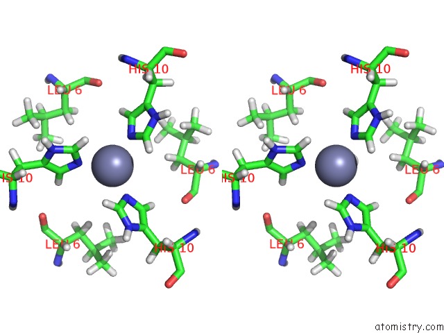
Stereo pair view
Mono view
Stereo pair view
A full contact list of Zinc with other atoms in the Zn binding
site number 2 of R6 Human Insulin Hexamer (Symmetric), uc(Nmr), 10 Structures within 5.0Å range:
|
Reference:
X.Chang,
A.M.Jorgensen,
P.Bardrum,
J.J.Led.
Solution Structures of the R6 Human Insulin Hexamer. Biochemistry V. 36 9409 1997.
ISSN: ISSN 0006-2960
PubMed: 9235985
DOI: 10.1021/BI9631069
Page generated: Sat Oct 12 22:03:16 2024
ISSN: ISSN 0006-2960
PubMed: 9235985
DOI: 10.1021/BI9631069
Last articles
Mn in 4R1PMn in 4R43
Mn in 4QSH
Mn in 4QSK
Mn in 4QTG
Mn in 4QNK
Mn in 4QRO
Mn in 4QSF
Mn in 4QS5
Mn in 4QRN