Zinc in PDB 8x44: Crystal Structure of DIMT1 in Complex with 5'-Methylthioadenosine From Pyrococcus Horikoshii (Formi)
Enzymatic activity of Crystal Structure of DIMT1 in Complex with 5'-Methylthioadenosine From Pyrococcus Horikoshii (Formi)
All present enzymatic activity of Crystal Structure of DIMT1 in Complex with 5'-Methylthioadenosine From Pyrococcus Horikoshii (Formi):
2.1.1.182;
2.1.1.182;
Protein crystallography data
The structure of Crystal Structure of DIMT1 in Complex with 5'-Methylthioadenosine From Pyrococcus Horikoshii (Formi), PDB code: 8x44
was solved by
S.Saha,
S.P.Kanaujia,
with X-Ray Crystallography technique. A brief refinement statistics is given in the table below:
| Resolution Low / High (Å) | 66.76 / 2.60 |
| Space group | P 2 21 21 |
| Cell size a, b, c (Å), α, β, γ (°) | 79.78, 85.34, 106.81, 90, 90, 90 |
| R / Rfree (%) | 20.5 / 27.3 |
Other elements in 8x44:
The structure of Crystal Structure of DIMT1 in Complex with 5'-Methylthioadenosine From Pyrococcus Horikoshii (Formi) also contains other interesting chemical elements:
| Chlorine | (Cl) | 1 atom |
Zinc Binding Sites:
The binding sites of Zinc atom in the Crystal Structure of DIMT1 in Complex with 5'-Methylthioadenosine From Pyrococcus Horikoshii (Formi)
(pdb code 8x44). This binding sites where shown within
5.0 Angstroms radius around Zinc atom.
In total 8 binding sites of Zinc where determined in the Crystal Structure of DIMT1 in Complex with 5'-Methylthioadenosine From Pyrococcus Horikoshii (Formi), PDB code: 8x44:
Jump to Zinc binding site number: 1; 2; 3; 4; 5; 6; 7; 8;
In total 8 binding sites of Zinc where determined in the Crystal Structure of DIMT1 in Complex with 5'-Methylthioadenosine From Pyrococcus Horikoshii (Formi), PDB code: 8x44:
Jump to Zinc binding site number: 1; 2; 3; 4; 5; 6; 7; 8;
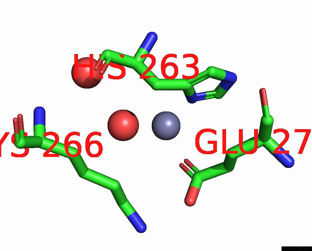
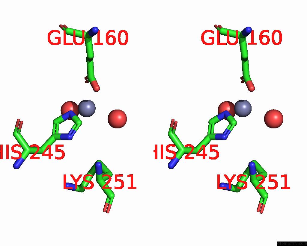
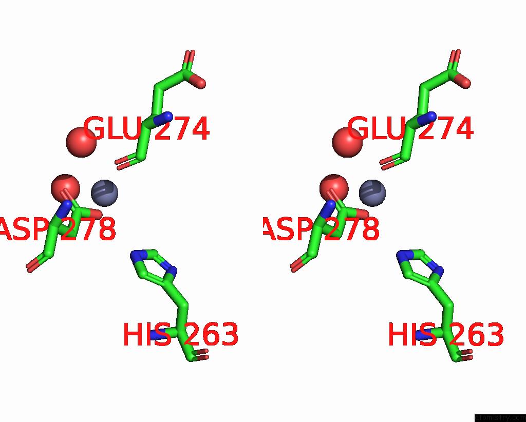
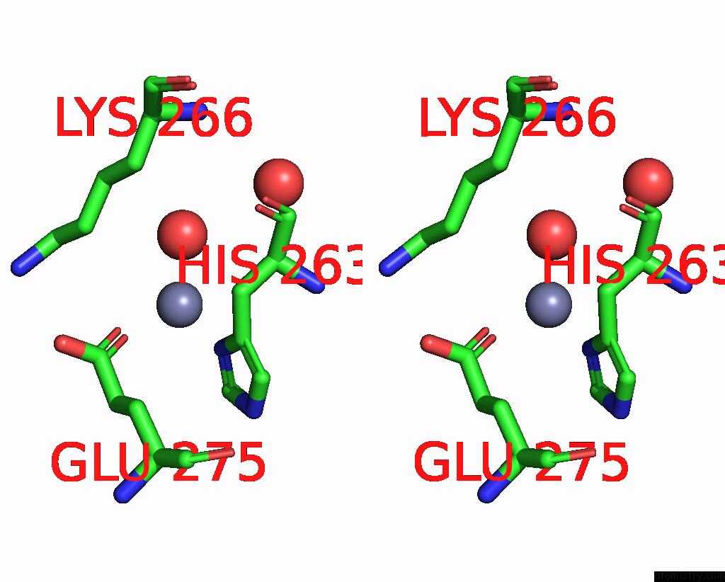
Zinc binding site 1 out of 8 in 8x44
Go back to
Zinc binding site 1 out
of 8 in the Crystal Structure of DIMT1 in Complex with 5'-Methylthioadenosine From Pyrococcus Horikoshii (Formi)
Mono view
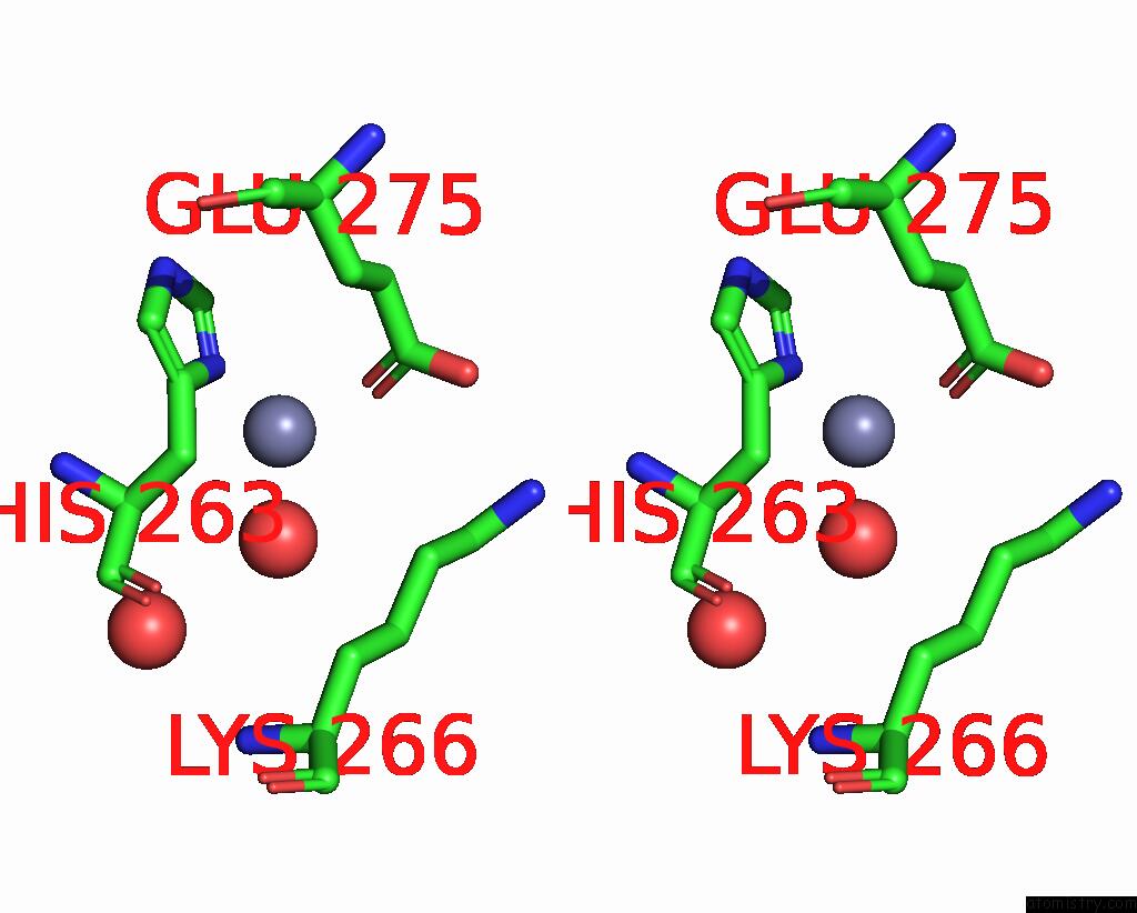
Stereo pair view
Mono view
Stereo pair view
A full contact list of Zinc with other atoms in the Zn binding
site number 1 of Crystal Structure of DIMT1 in Complex with 5'-Methylthioadenosine From Pyrococcus Horikoshii (Formi) within 5.0Å range:
|
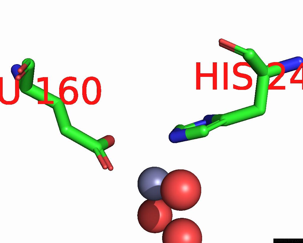
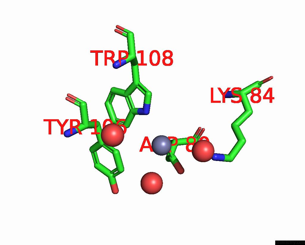
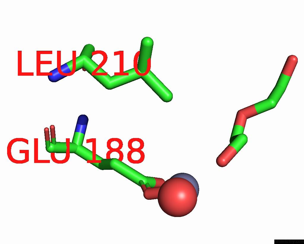
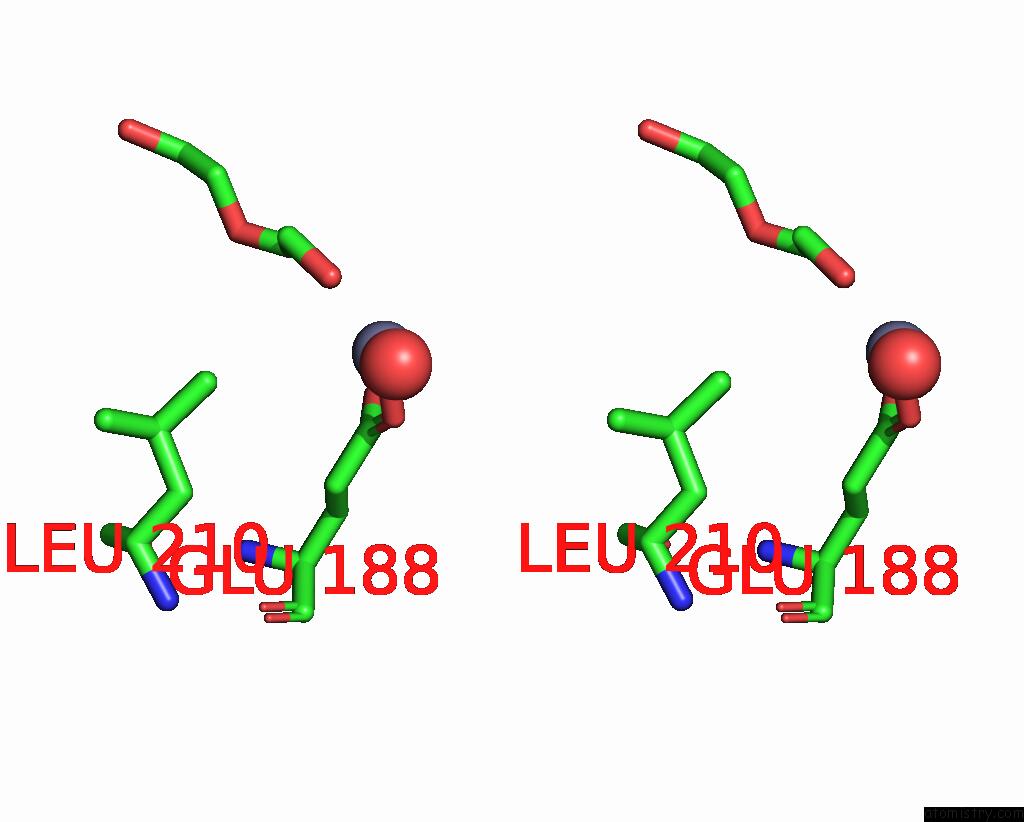
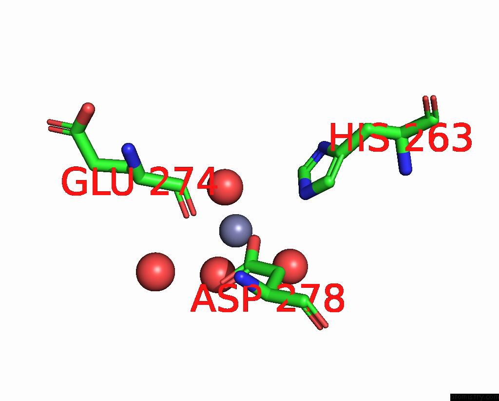
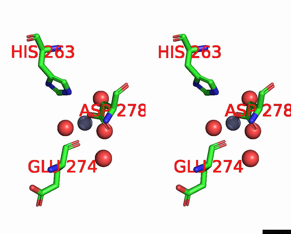
Zinc binding site 2 out of 8 in 8x44
Go back to
Zinc binding site 2 out
of 8 in the Crystal Structure of DIMT1 in Complex with 5'-Methylthioadenosine From Pyrococcus Horikoshii (Formi)
Mono view

Stereo pair view
Mono view
Stereo pair view
A full contact list of Zinc with other atoms in the Zn binding
site number 2 of Crystal Structure of DIMT1 in Complex with 5'-Methylthioadenosine From Pyrococcus Horikoshii (Formi) within 5.0Å range:
|
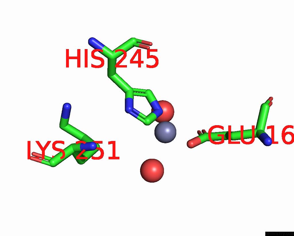
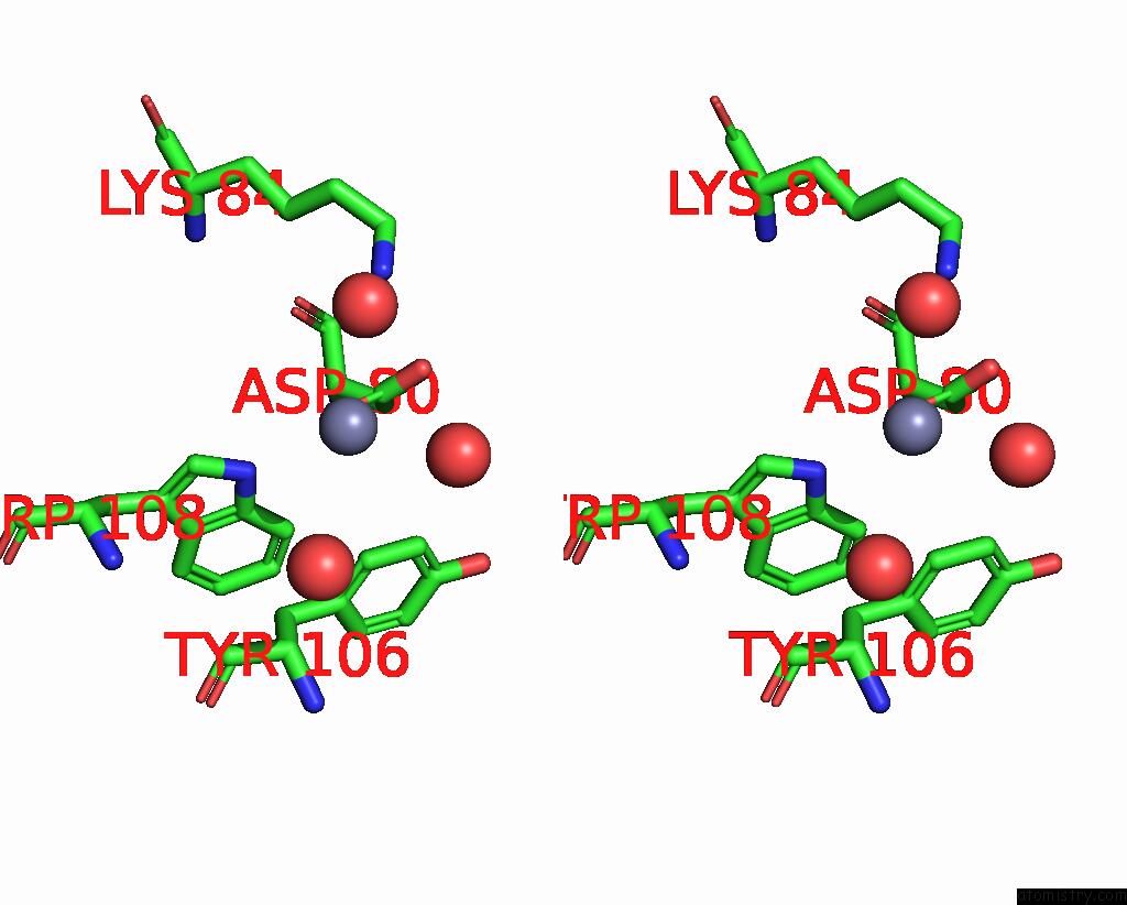
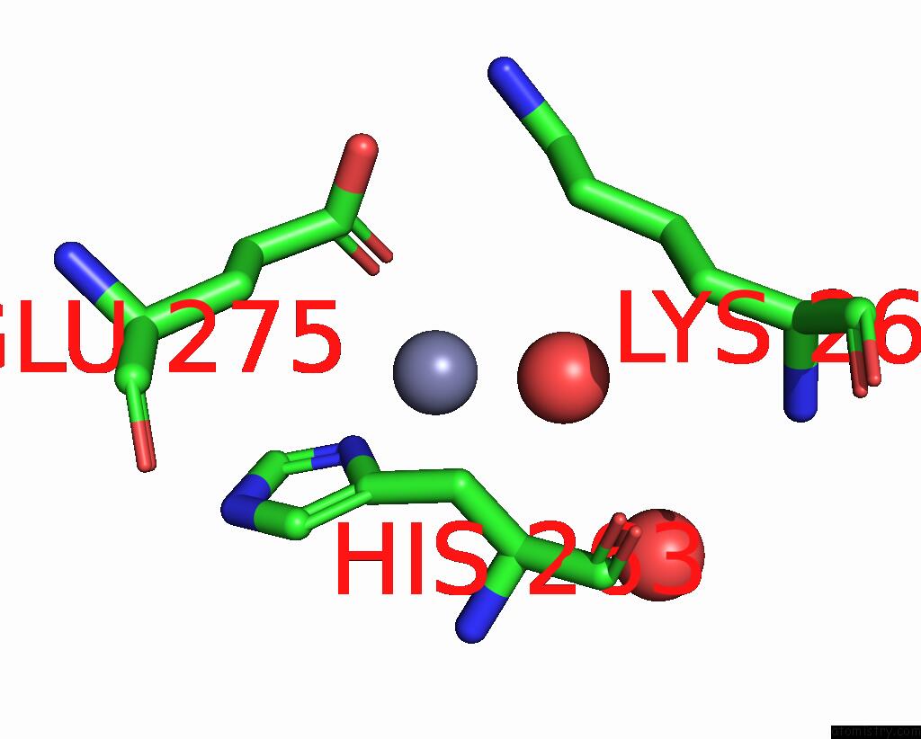
Zinc binding site 3 out of 8 in 8x44
Go back to
Zinc binding site 3 out
of 8 in the Crystal Structure of DIMT1 in Complex with 5'-Methylthioadenosine From Pyrococcus Horikoshii (Formi)
Mono view
Stereo pair view
Mono view
Stereo pair view
A full contact list of Zinc with other atoms in the Zn binding
site number 3 of Crystal Structure of DIMT1 in Complex with 5'-Methylthioadenosine From Pyrococcus Horikoshii (Formi) within 5.0Å range:
|
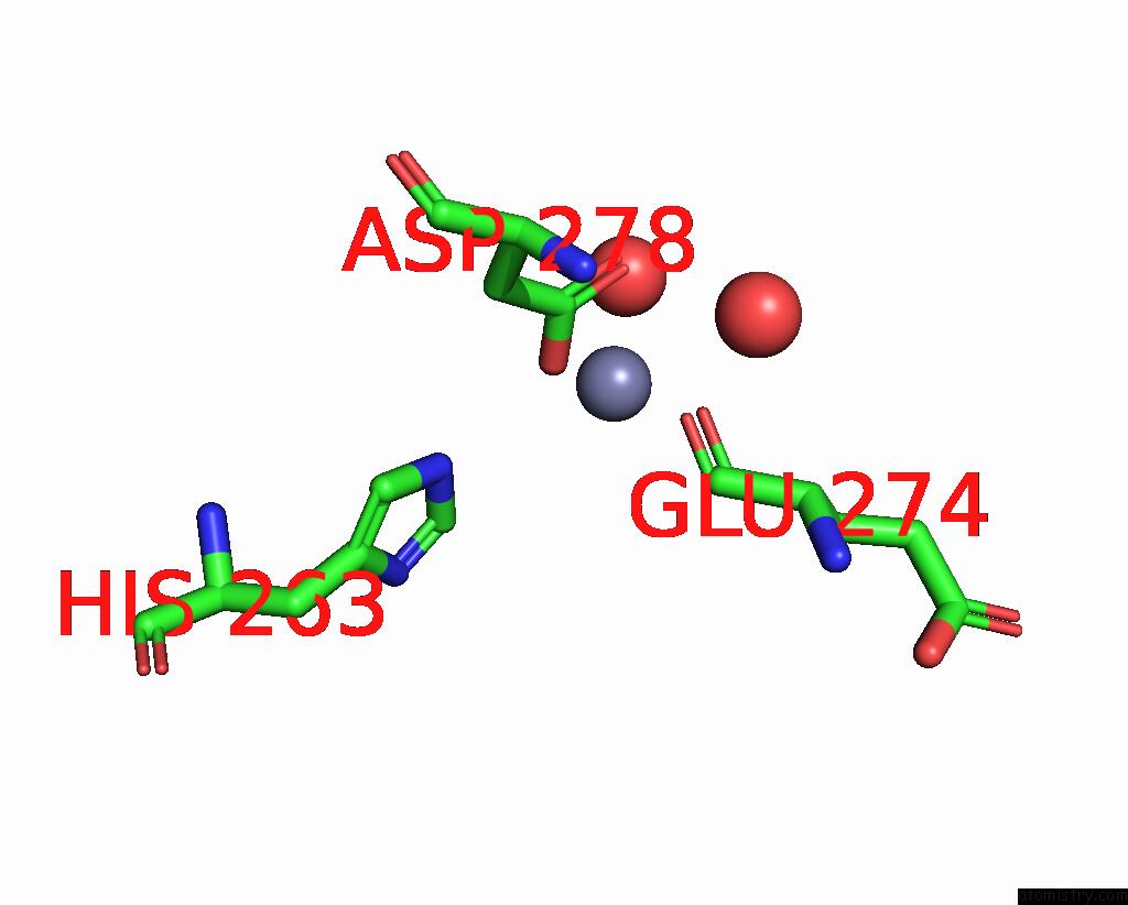
Zinc binding site 4 out of 8 in 8x44
Go back to
Zinc binding site 4 out
of 8 in the Crystal Structure of DIMT1 in Complex with 5'-Methylthioadenosine From Pyrococcus Horikoshii (Formi)
Mono view
Stereo pair view
Mono view
Stereo pair view
A full contact list of Zinc with other atoms in the Zn binding
site number 4 of Crystal Structure of DIMT1 in Complex with 5'-Methylthioadenosine From Pyrococcus Horikoshii (Formi) within 5.0Å range:
|
Zinc binding site 5 out of 8 in 8x44
Go back to
Zinc binding site 5 out
of 8 in the Crystal Structure of DIMT1 in Complex with 5'-Methylthioadenosine From Pyrococcus Horikoshii (Formi)
Mono view
Stereo pair view
Mono view
Stereo pair view
A full contact list of Zinc with other atoms in the Zn binding
site number 5 of Crystal Structure of DIMT1 in Complex with 5'-Methylthioadenosine From Pyrococcus Horikoshii (Formi) within 5.0Å range:
|
Zinc binding site 6 out of 8 in 8x44
Go back to
Zinc binding site 6 out
of 8 in the Crystal Structure of DIMT1 in Complex with 5'-Methylthioadenosine From Pyrococcus Horikoshii (Formi)
Mono view
Stereo pair view
Mono view
Stereo pair view
A full contact list of Zinc with other atoms in the Zn binding
site number 6 of Crystal Structure of DIMT1 in Complex with 5'-Methylthioadenosine From Pyrococcus Horikoshii (Formi) within 5.0Å range:
|
Zinc binding site 7 out of 8 in 8x44
Go back to
Zinc binding site 7 out
of 8 in the Crystal Structure of DIMT1 in Complex with 5'-Methylthioadenosine From Pyrococcus Horikoshii (Formi)
Mono view
Stereo pair view
Mono view
Stereo pair view
A full contact list of Zinc with other atoms in the Zn binding
site number 7 of Crystal Structure of DIMT1 in Complex with 5'-Methylthioadenosine From Pyrococcus Horikoshii (Formi) within 5.0Å range:
|
Zinc binding site 8 out of 8 in 8x44
Go back to
Zinc binding site 8 out
of 8 in the Crystal Structure of DIMT1 in Complex with 5'-Methylthioadenosine From Pyrococcus Horikoshii (Formi)
Mono view
Stereo pair view
Mono view
Stereo pair view
A full contact list of Zinc with other atoms in the Zn binding
site number 8 of Crystal Structure of DIMT1 in Complex with 5'-Methylthioadenosine From Pyrococcus Horikoshii (Formi) within 5.0Å range:
|
Reference:
S.Saha,
S.P.Kanaujia.
Structural and Functional Characterization of Archaeal DIMT1 Unveils Distinct Protein Dynamics Essential For Efficient Catalysis. Structure 2024.
ISSN: ISSN 0969-2126
PubMed: 39146930
DOI: 10.1016/J.STR.2024.07.013
Page generated: Thu Oct 31 13:45:23 2024
ISSN: ISSN 0969-2126
PubMed: 39146930
DOI: 10.1016/J.STR.2024.07.013
Last articles
Zn in 9J0NZn in 9J0O
Zn in 9J0P
Zn in 9FJX
Zn in 9EKB
Zn in 9C0F
Zn in 9CAH
Zn in 9CH0
Zn in 9CH3
Zn in 9CH1