Zinc in PDB 8tvc: Crystal Structure of RA3G-Ssdna-Aa
Protein crystallography data
The structure of Crystal Structure of RA3G-Ssdna-Aa, PDB code: 8tvc
was solved by
H.Yang,
J.I.Pacheco,
X.S.Chen,
with X-Ray Crystallography technique. A brief refinement statistics is given in the table below:
| Resolution Low / High (Å) | 43.08 / 1.93 |
| Space group | P 21 21 21 |
| Cell size a, b, c (Å), α, β, γ (°) | 55.429, 68.461, 126.735, 90, 90, 90 |
| R / Rfree (%) | 17.8 / 20.8 |
Zinc Binding Sites:
The binding sites of Zinc atom in the Crystal Structure of RA3G-Ssdna-Aa
(pdb code 8tvc). This binding sites where shown within
5.0 Angstroms radius around Zinc atom.
In total 2 binding sites of Zinc where determined in the Crystal Structure of RA3G-Ssdna-Aa, PDB code: 8tvc:
Jump to Zinc binding site number: 1; 2;
In total 2 binding sites of Zinc where determined in the Crystal Structure of RA3G-Ssdna-Aa, PDB code: 8tvc:
Jump to Zinc binding site number: 1; 2;
Zinc binding site 1 out of 2 in 8tvc
Go back to
Zinc binding site 1 out
of 2 in the Crystal Structure of RA3G-Ssdna-Aa
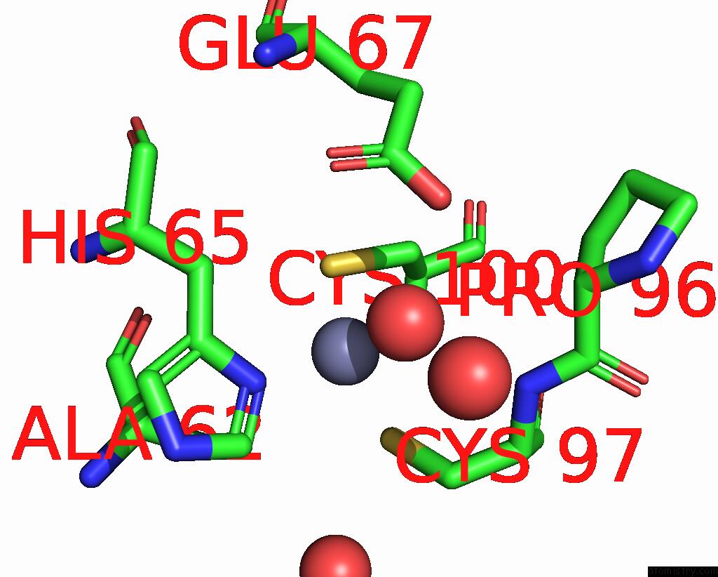
Mono view
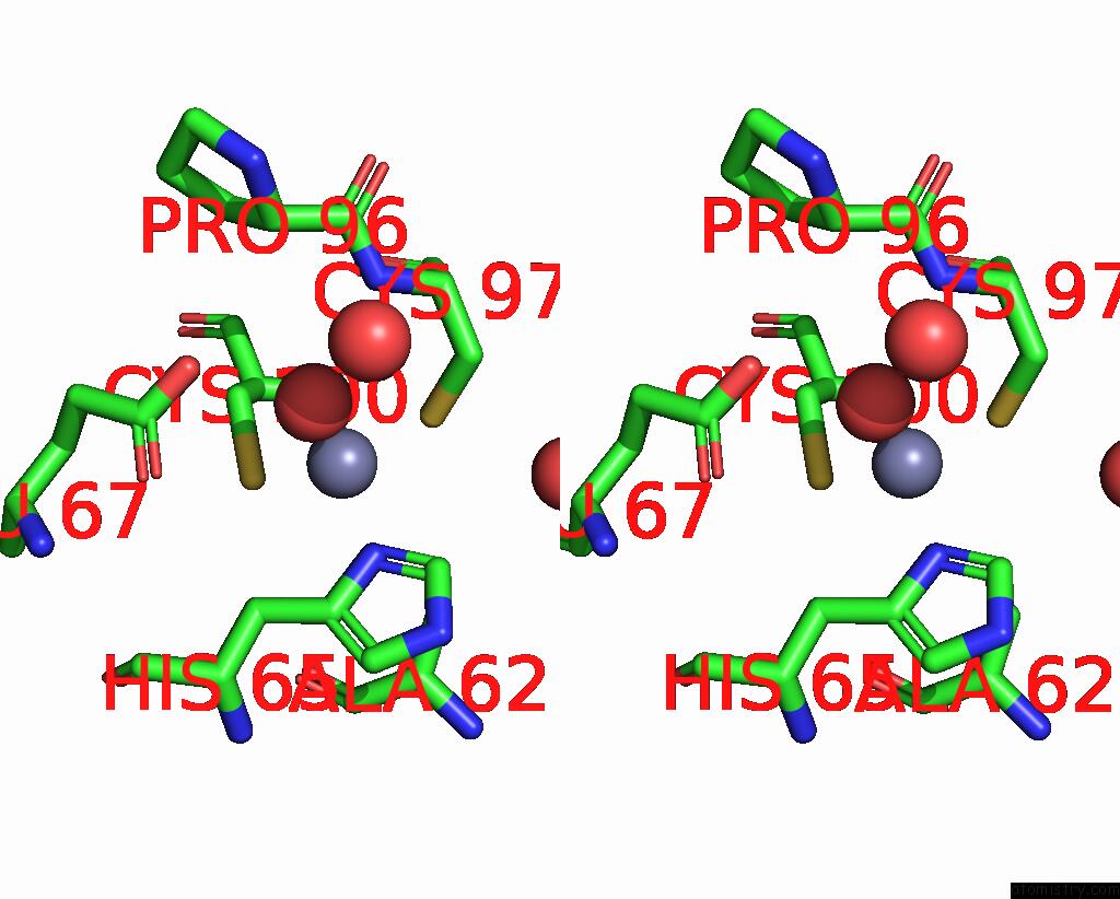
Stereo pair view
Mono view
Stereo pair view
A full contact list of Zinc with other atoms in the Zn binding
site number 1 of Crystal Structure of RA3G-Ssdna-Aa within 5.0Å range:
|
Zinc binding site 2 out of 2 in 8tvc
Go back to
Zinc binding site 2 out
of 2 in the Crystal Structure of RA3G-Ssdna-Aa
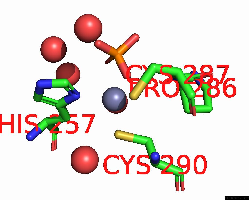
Mono view
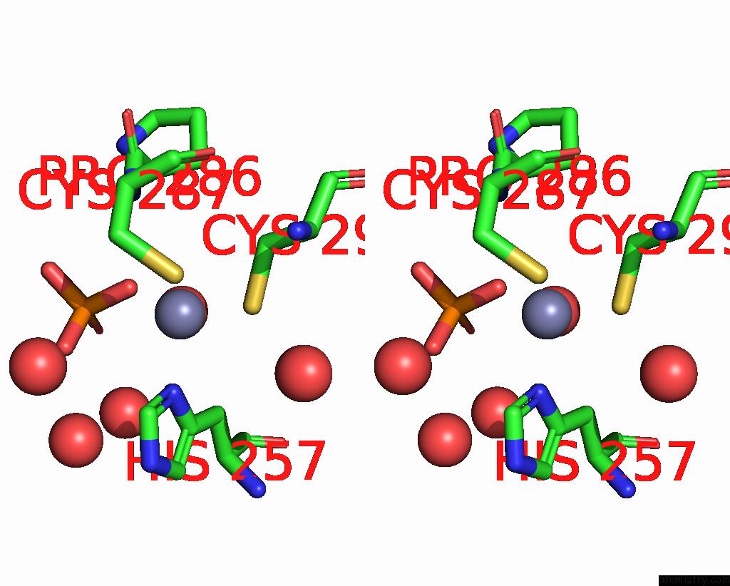
Stereo pair view
Mono view
Stereo pair view
A full contact list of Zinc with other atoms in the Zn binding
site number 2 of Crystal Structure of RA3G-Ssdna-Aa within 5.0Å range:
|
Reference:
H.Yang,
J.Pacheco,
K.Kim,
A.Bokani,
F.Ito,
D.Ebrahimi,
X.S.Chen.
Molecular Mechanism For Regulating APOBEC3G Dna Editing Function By the Non-Catalytic Domain. Nat Commun V. 15 8773 2024.
ISSN: ESSN 2041-1723
PubMed: 39389938
DOI: 10.1038/S41467-024-52671-1
Page generated: Wed Nov 13 13:43:24 2024
ISSN: ESSN 2041-1723
PubMed: 39389938
DOI: 10.1038/S41467-024-52671-1
Last articles
Zn in 9J0NZn in 9J0O
Zn in 9J0P
Zn in 9FJX
Zn in 9EKB
Zn in 9C0F
Zn in 9CAH
Zn in 9CH0
Zn in 9CH3
Zn in 9CH1