Zinc in PDB 8szu: Structure of KDAC1-Citarinostat Complex From Acinetobacter Baumannii
Protein crystallography data
The structure of Structure of KDAC1-Citarinostat Complex From Acinetobacter Baumannii, PDB code: 8szu
was solved by
P.R.Watson,
D.W.Christianson,
with X-Ray Crystallography technique. A brief refinement statistics is given in the table below:
| Resolution Low / High (Å) | 43.73 / 1.75 |
| Space group | P 41 21 2 |
| Cell size a, b, c (Å), α, β, γ (°) | 80.594, 80.594, 204.645, 90, 90, 90 |
| R / Rfree (%) | 18.3 / 21.1 |
Other elements in 8szu:
The structure of Structure of KDAC1-Citarinostat Complex From Acinetobacter Baumannii also contains other interesting chemical elements:
| Potassium | (K) | 4 atoms |
| Chlorine | (Cl) | 4 atoms |
Zinc Binding Sites:
The binding sites of Zinc atom in the Structure of KDAC1-Citarinostat Complex From Acinetobacter Baumannii
(pdb code 8szu). This binding sites where shown within
5.0 Angstroms radius around Zinc atom.
In total 2 binding sites of Zinc where determined in the Structure of KDAC1-Citarinostat Complex From Acinetobacter Baumannii, PDB code: 8szu:
Jump to Zinc binding site number: 1; 2;
In total 2 binding sites of Zinc where determined in the Structure of KDAC1-Citarinostat Complex From Acinetobacter Baumannii, PDB code: 8szu:
Jump to Zinc binding site number: 1; 2;
Zinc binding site 1 out of 2 in 8szu
Go back to
Zinc binding site 1 out
of 2 in the Structure of KDAC1-Citarinostat Complex From Acinetobacter Baumannii
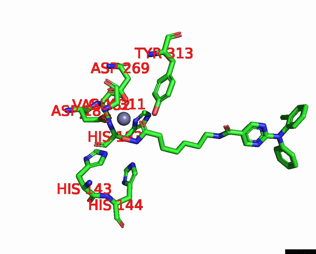
Mono view
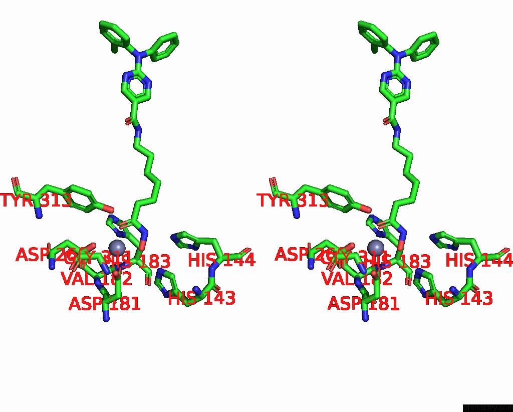
Stereo pair view
Mono view
Stereo pair view
A full contact list of Zinc with other atoms in the Zn binding
site number 1 of Structure of KDAC1-Citarinostat Complex From Acinetobacter Baumannii within 5.0Å range:
|
Zinc binding site 2 out of 2 in 8szu
Go back to
Zinc binding site 2 out
of 2 in the Structure of KDAC1-Citarinostat Complex From Acinetobacter Baumannii

Mono view
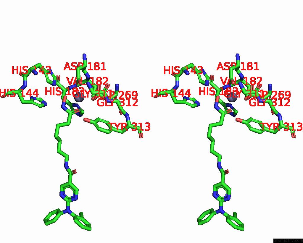
Stereo pair view
Mono view
Stereo pair view
A full contact list of Zinc with other atoms in the Zn binding
site number 2 of Structure of KDAC1-Citarinostat Complex From Acinetobacter Baumannii within 5.0Å range:
|
Reference:
P.R.Watson,
D.W.Christianson.
Structure and Function of KDAC1, A Class II Deacetylase From the Multidrug-Resistant Pathogen Acinetobacter Baumannii. Biochemistry 2023.
ISSN: ISSN 0006-2960
PubMed: 37624144
DOI: 10.1021/ACS.BIOCHEM.3C00288
Page generated: Thu Oct 31 11:20:04 2024
ISSN: ISSN 0006-2960
PubMed: 37624144
DOI: 10.1021/ACS.BIOCHEM.3C00288
Last articles
Zn in 9J0NZn in 9J0O
Zn in 9J0P
Zn in 9FJX
Zn in 9EKB
Zn in 9C0F
Zn in 9CAH
Zn in 9CH0
Zn in 9CH3
Zn in 9CH1