Zinc in PDB 8rww: Crystal Structure of Native Alpha-Keto C-Methyl Transferase Sgvm Bound to Ketoleucine
Protein crystallography data
The structure of Crystal Structure of Native Alpha-Keto C-Methyl Transferase Sgvm Bound to Ketoleucine, PDB code: 8rww
was solved by
S.Gerhardt,
J.N.Andexer,
with X-Ray Crystallography technique. A brief refinement statistics is given in the table below:
| Resolution Low / High (Å) | 20.09 / 1.95 |
| Space group | P 43 21 2 |
| Cell size a, b, c (Å), α, β, γ (°) | 67.278, 67.278, 182.967, 90, 90, 90 |
| R / Rfree (%) | 18.2 / 21.2 |
Other elements in 8rww:
The structure of Crystal Structure of Native Alpha-Keto C-Methyl Transferase Sgvm Bound to Ketoleucine also contains other interesting chemical elements:
| Chlorine | (Cl) | 1 atom |
Zinc Binding Sites:
The binding sites of Zinc atom in the Crystal Structure of Native Alpha-Keto C-Methyl Transferase Sgvm Bound to Ketoleucine
(pdb code 8rww). This binding sites where shown within
5.0 Angstroms radius around Zinc atom.
In total only one binding site of Zinc was determined in the Crystal Structure of Native Alpha-Keto C-Methyl Transferase Sgvm Bound to Ketoleucine, PDB code: 8rww:
In total only one binding site of Zinc was determined in the Crystal Structure of Native Alpha-Keto C-Methyl Transferase Sgvm Bound to Ketoleucine, PDB code: 8rww:
Zinc binding site 1 out of 1 in 8rww
Go back to
Zinc binding site 1 out
of 1 in the Crystal Structure of Native Alpha-Keto C-Methyl Transferase Sgvm Bound to Ketoleucine
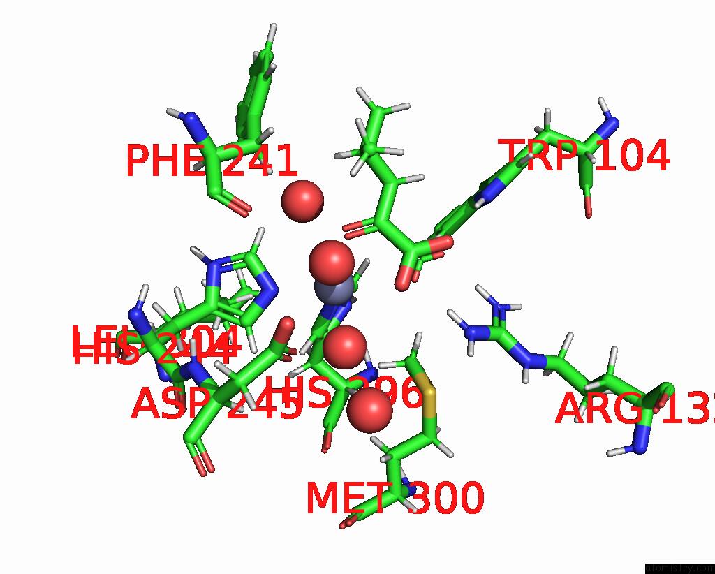
Mono view
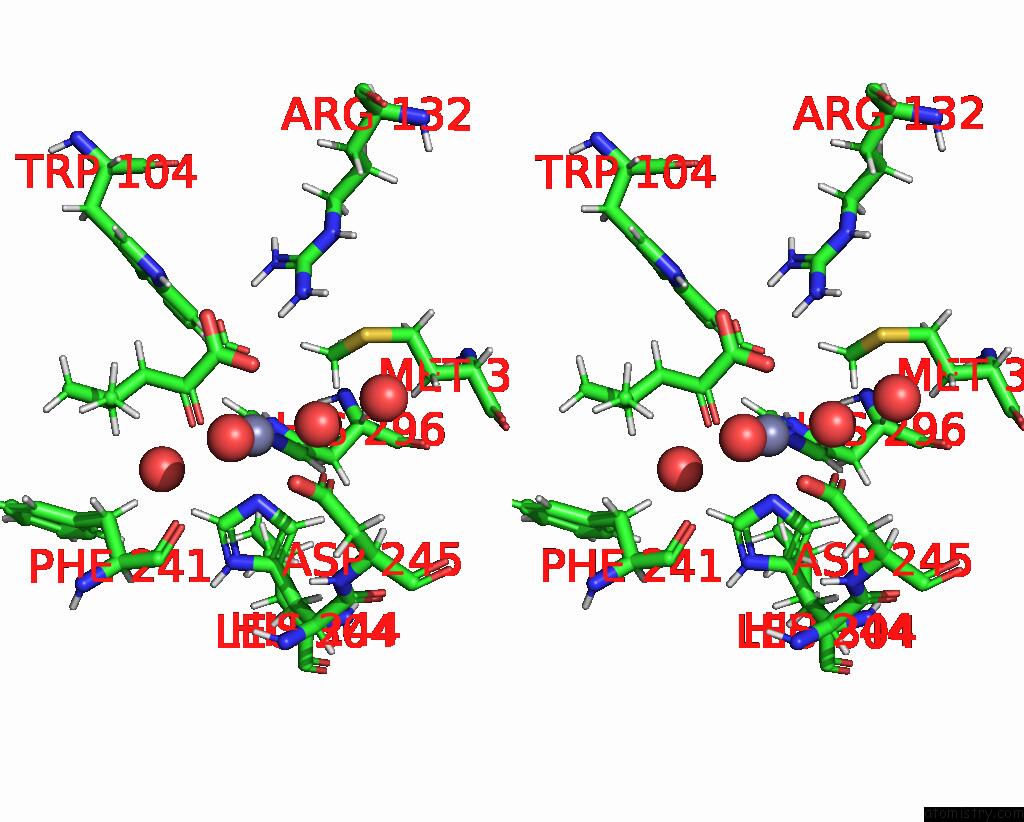
Stereo pair view
Mono view
Stereo pair view
A full contact list of Zinc with other atoms in the Zn binding
site number 1 of Crystal Structure of Native Alpha-Keto C-Methyl Transferase Sgvm Bound to Ketoleucine within 5.0Å range:
|
Reference:
C.Sommer-Kamann,
J.Breiltgens,
Z.Zou,
S.Gerhardt,
R.Saleem-Batcha,
F.Kemper,
O.Einsle,
J.N.Andexer,
M.Muller.
Structures and Protein Engineering of the Alpha-Keto Acid C-Methyltransferases Sgvm and Mrsa For Rational Substrate Transfer. Chembiochem 00258 2024.
ISSN: ESSN 1439-7633
PubMed: 38887142
DOI: 10.1002/CBIC.202400258
Page generated: Thu Oct 31 10:46:31 2024
ISSN: ESSN 1439-7633
PubMed: 38887142
DOI: 10.1002/CBIC.202400258
Last articles
Zn in 9J0NZn in 9J0O
Zn in 9J0P
Zn in 9FJX
Zn in 9EKB
Zn in 9C0F
Zn in 9CAH
Zn in 9CH0
Zn in 9CH3
Zn in 9CH1