Zinc in PDB 8ruf: Crystal Structure of Rhizobium Etli L-Asparaginase Reav D187A Mutant
Protein crystallography data
The structure of Crystal Structure of Rhizobium Etli L-Asparaginase Reav D187A Mutant, PDB code: 8ruf
was solved by
K.Pokrywka,
M.Grzechowiak,
J.Sliwiak,
P.Worsztynowicz,
J.I.Loch,
M.Ruszkowski,
M.Gilski,
M.Jaskolski,
with X-Ray Crystallography technique. A brief refinement statistics is given in the table below:
Resolution Low / High (Å)
|
48.57 /
1.60
|
Space group
|
P 1 21 1
|
Cell size a, b, c (Å), α, β, γ (°)
|
77.715,
91.26,
114.197,
90,
96.95,
90
|
R / Rfree (%)
|
15.8 /
18.4
|
Other elements in 8ruf:
The structure of Crystal Structure of Rhizobium Etli L-Asparaginase Reav D187A Mutant also contains other interesting chemical elements:
Zinc Binding Sites:
The binding sites of Zinc atom in the Crystal Structure of Rhizobium Etli L-Asparaginase Reav D187A Mutant
(pdb code 8ruf). This binding sites where shown within
5.0 Angstroms radius around Zinc atom.
In total 4 binding sites of Zinc where determined in the
Crystal Structure of Rhizobium Etli L-Asparaginase Reav D187A Mutant, PDB code: 8ruf:
Jump to Zinc binding site number:
1;
2;
3;
4;
Zinc binding site 1 out
of 4 in 8ruf
Go back to
Zinc Binding Sites List in 8ruf
Zinc binding site 1 out
of 4 in the Crystal Structure of Rhizobium Etli L-Asparaginase Reav D187A Mutant
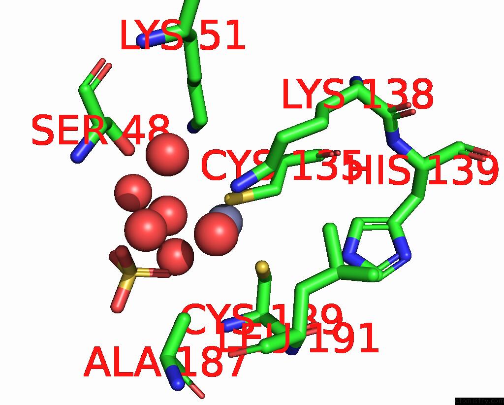
Mono view
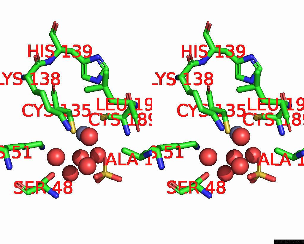
Stereo pair view
|
A full contact list of Zinc with other atoms in the Zn binding
site number 1 of Crystal Structure of Rhizobium Etli L-Asparaginase Reav D187A Mutant within 5.0Å range:
probe
|
atom
|
residue
|
distance (Å)
|
B
|
Occ
|
A:Zn404
b:21.5
occ:0.50
|
O
|
A:HOH502
|
2.2
|
30.4
|
0.5
|
NZ
|
A:LYS138
|
2.2
|
22.4
|
1.0
|
SG
|
A:CYS189
|
2.3
|
27.3
|
1.0
|
SG
|
A:CYS135
|
2.3
|
22.0
|
1.0
|
CB
|
A:CYS189
|
3.0
|
27.9
|
1.0
|
CE
|
A:LYS138
|
3.1
|
19.7
|
1.0
|
O1
|
A:SO4401
|
3.1
|
27.4
|
1.0
|
CB
|
A:CYS135
|
3.3
|
18.4
|
1.0
|
O
|
A:HOH532
|
4.0
|
22.8
|
1.0
|
O3
|
A:SO4401
|
4.1
|
39.7
|
1.0
|
CE
|
A:LYS51
|
4.1
|
20.0
|
1.0
|
S
|
A:SO4401
|
4.1
|
35.6
|
1.0
|
O
|
A:HOH681
|
4.2
|
20.0
|
1.0
|
O
|
A:HOH528
|
4.2
|
34.2
|
1.0
|
CA
|
A:CYS189
|
4.2
|
23.5
|
1.0
|
N
|
A:CYS189
|
4.3
|
21.8
|
1.0
|
CB
|
A:ALA187
|
4.3
|
23.9
|
1.0
|
O
|
A:HOH564
|
4.4
|
23.5
|
1.0
|
NZ
|
A:LYS51
|
4.5
|
21.1
|
1.0
|
CD
|
A:LYS138
|
4.5
|
19.4
|
1.0
|
CA
|
A:CYS135
|
4.7
|
18.2
|
1.0
|
O2
|
A:SO4401
|
4.8
|
42.6
|
1.0
|
NE2
|
A:HIS139
|
4.9
|
17.0
|
1.0
|
CD1
|
A:LEU191
|
4.9
|
20.5
|
1.0
|
OG
|
A:SER48
|
4.9
|
25.7
|
1.0
|
O
|
A:HOH508
|
5.0
|
28.3
|
1.0
|
C
|
A:CYS189
|
5.0
|
26.9
|
1.0
|
|
Zinc binding site 2 out
of 4 in 8ruf
Go back to
Zinc Binding Sites List in 8ruf
Zinc binding site 2 out
of 4 in the Crystal Structure of Rhizobium Etli L-Asparaginase Reav D187A Mutant
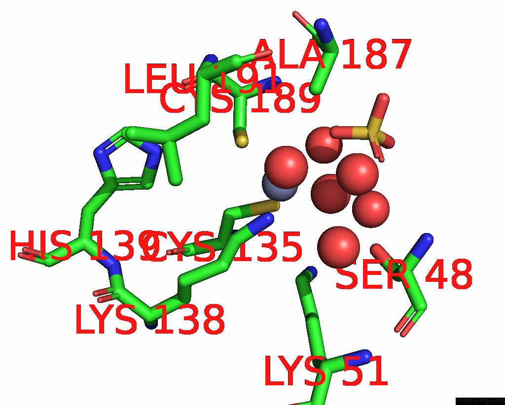
Mono view
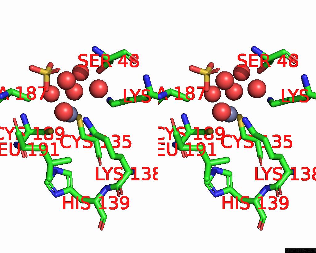
Stereo pair view
|
A full contact list of Zinc with other atoms in the Zn binding
site number 2 of Crystal Structure of Rhizobium Etli L-Asparaginase Reav D187A Mutant within 5.0Å range:
probe
|
atom
|
residue
|
distance (Å)
|
B
|
Occ
|
B:Zn404
b:20.6
occ:0.50
|
O
|
B:HOH507
|
2.0
|
23.4
|
0.5
|
NZ
|
B:LYS138
|
2.2
|
19.5
|
1.0
|
SG
|
B:CYS189
|
2.2
|
25.5
|
1.0
|
SG
|
B:CYS135
|
2.3
|
21.4
|
1.0
|
CB
|
B:CYS189
|
2.9
|
26.4
|
1.0
|
CE
|
B:LYS138
|
3.1
|
18.8
|
1.0
|
O2
|
B:SO4401
|
3.1
|
27.4
|
1.0
|
CB
|
B:CYS135
|
3.3
|
14.8
|
1.0
|
O4
|
B:SO4401
|
4.0
|
29.9
|
1.0
|
CE
|
B:LYS51
|
4.0
|
17.5
|
1.0
|
O
|
B:HOH531
|
4.1
|
19.4
|
1.0
|
O
|
B:HOH723
|
4.1
|
17.7
|
1.0
|
S
|
B:SO4401
|
4.1
|
30.6
|
1.0
|
O
|
B:HOH519
|
4.2
|
30.9
|
1.0
|
CA
|
B:CYS189
|
4.2
|
23.6
|
1.0
|
CB
|
B:ALA187
|
4.3
|
17.8
|
1.0
|
N
|
B:CYS189
|
4.3
|
22.6
|
1.0
|
NZ
|
B:LYS51
|
4.3
|
17.3
|
1.0
|
O
|
B:HOH550
|
4.4
|
21.3
|
1.0
|
CD
|
B:LYS138
|
4.5
|
17.9
|
1.0
|
CA
|
B:CYS135
|
4.7
|
15.6
|
1.0
|
OG
|
B:SER48
|
4.9
|
22.0
|
1.0
|
O
|
B:HOH503
|
4.9
|
27.3
|
1.0
|
CD1
|
B:LEU191
|
5.0
|
20.3
|
1.0
|
C
|
B:CYS189
|
5.0
|
25.4
|
1.0
|
O1
|
B:SO4401
|
5.0
|
34.9
|
1.0
|
NE2
|
B:HIS139
|
5.0
|
17.4
|
1.0
|
|
Zinc binding site 3 out
of 4 in 8ruf
Go back to
Zinc Binding Sites List in 8ruf
Zinc binding site 3 out
of 4 in the Crystal Structure of Rhizobium Etli L-Asparaginase Reav D187A Mutant
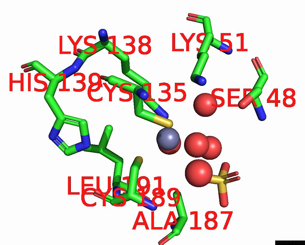
Mono view
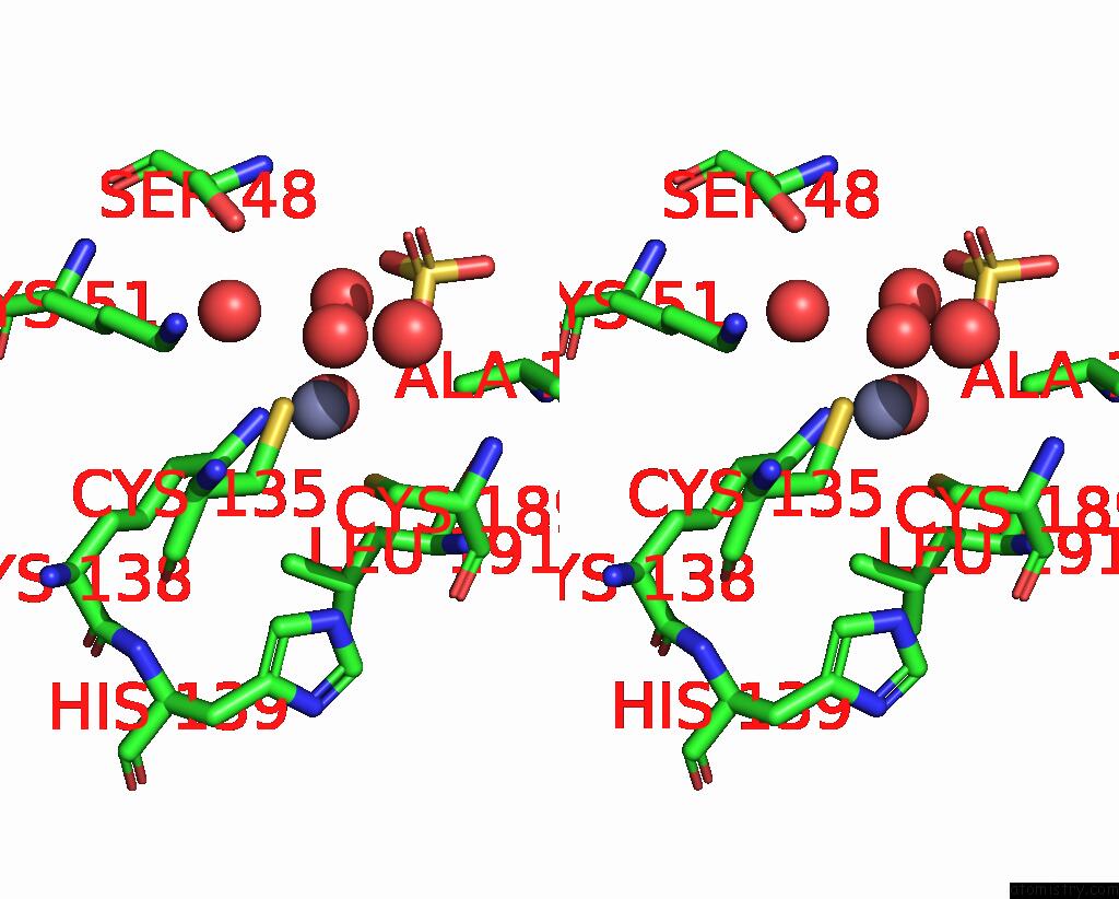
Stereo pair view
|
A full contact list of Zinc with other atoms in the Zn binding
site number 3 of Crystal Structure of Rhizobium Etli L-Asparaginase Reav D187A Mutant within 5.0Å range:
probe
|
atom
|
residue
|
distance (Å)
|
B
|
Occ
|
C:Zn403
b:19.9
occ:0.50
|
O
|
C:HOH501
|
1.9
|
25.0
|
0.5
|
NZ
|
C:LYS138
|
2.2
|
21.8
|
1.0
|
SG
|
C:CYS189
|
2.3
|
25.3
|
1.0
|
SG
|
C:CYS135
|
2.3
|
23.8
|
1.0
|
CB
|
C:CYS189
|
2.9
|
24.4
|
1.0
|
O3
|
C:SO4401
|
3.0
|
28.8
|
1.0
|
CE
|
C:LYS138
|
3.1
|
21.5
|
1.0
|
CB
|
C:CYS135
|
3.3
|
17.6
|
1.0
|
O1
|
C:SO4401
|
4.0
|
35.4
|
1.0
|
CE
|
C:LYS51
|
4.0
|
17.6
|
1.0
|
O
|
C:HOH529
|
4.0
|
23.3
|
1.0
|
S
|
C:SO4401
|
4.0
|
35.7
|
1.0
|
O
|
C:HOH709
|
4.1
|
17.5
|
1.0
|
CA
|
C:CYS189
|
4.2
|
26.9
|
1.0
|
CB
|
C:ALA187
|
4.2
|
21.7
|
1.0
|
O
|
C:HOH531
|
4.2
|
35.8
|
1.0
|
N
|
C:CYS189
|
4.2
|
22.1
|
1.0
|
NZ
|
C:LYS51
|
4.4
|
18.6
|
1.0
|
O
|
C:HOH537
|
4.4
|
20.2
|
1.0
|
CD
|
C:LYS138
|
4.5
|
17.7
|
1.0
|
CA
|
C:CYS135
|
4.6
|
15.4
|
1.0
|
O4
|
C:SO4401
|
4.8
|
40.1
|
1.0
|
C
|
C:CYS189
|
4.9
|
28.7
|
1.0
|
NE2
|
C:HIS139
|
5.0
|
18.5
|
1.0
|
OG
|
C:SER48
|
5.0
|
26.1
|
1.0
|
CD1
|
C:LEU191
|
5.0
|
19.3
|
1.0
|
|
Zinc binding site 4 out
of 4 in 8ruf
Go back to
Zinc Binding Sites List in 8ruf
Zinc binding site 4 out
of 4 in the Crystal Structure of Rhizobium Etli L-Asparaginase Reav D187A Mutant
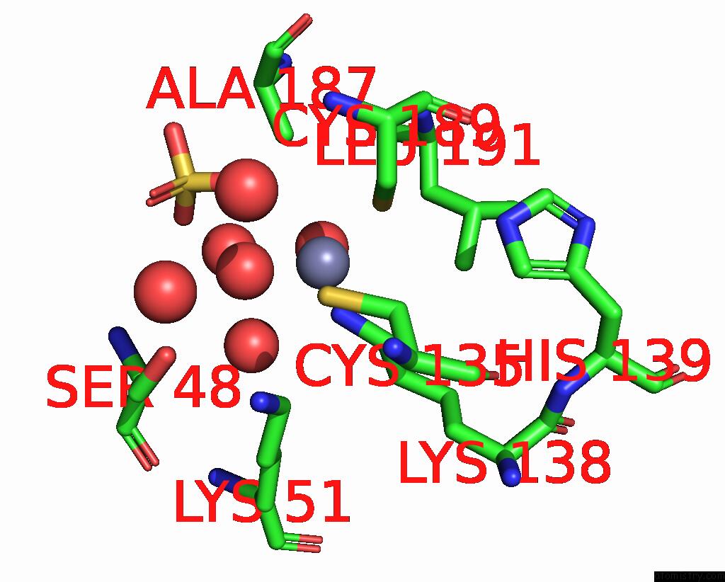
Mono view
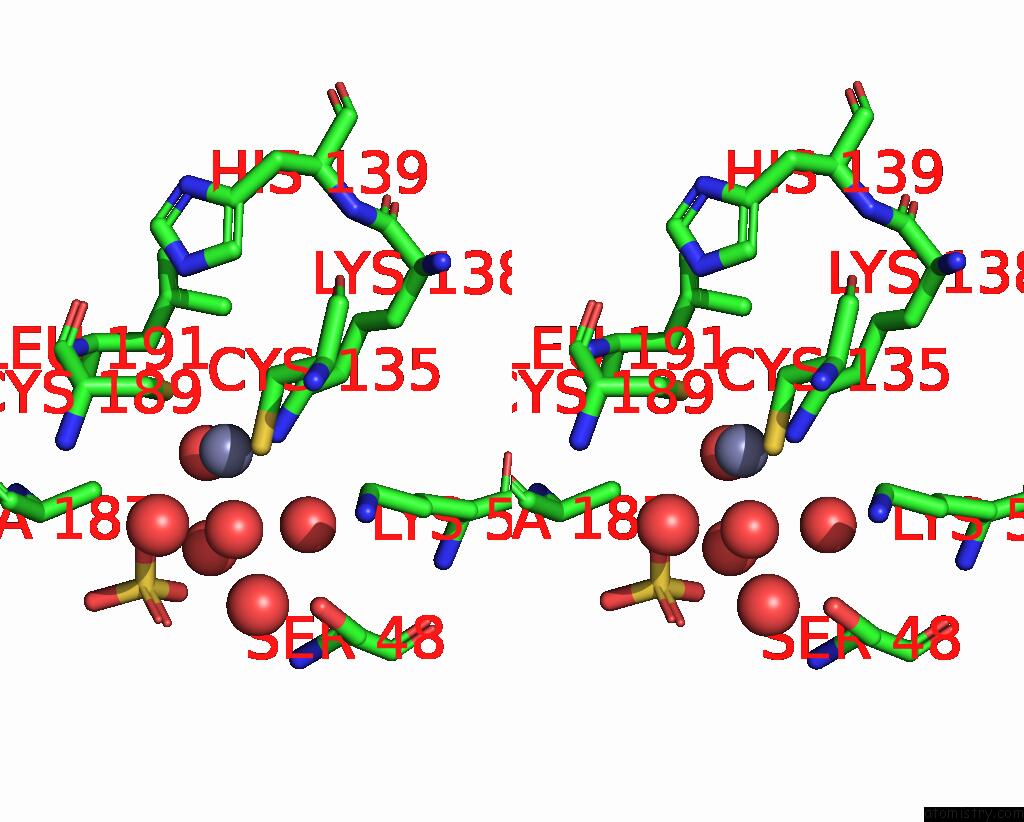
Stereo pair view
|
A full contact list of Zinc with other atoms in the Zn binding
site number 4 of Crystal Structure of Rhizobium Etli L-Asparaginase Reav D187A Mutant within 5.0Å range:
probe
|
atom
|
residue
|
distance (Å)
|
B
|
Occ
|
D:Zn408
b:20.1
occ:0.50
|
O
|
D:HOH503
|
2.1
|
26.4
|
0.5
|
NZ
|
D:LYS138
|
2.2
|
19.2
|
1.0
|
SG
|
D:CYS189
|
2.3
|
25.7
|
1.0
|
SG
|
D:CYS135
|
2.3
|
22.7
|
1.0
|
CB
|
D:CYS189
|
3.0
|
28.2
|
1.0
|
CE
|
D:LYS138
|
3.0
|
20.4
|
1.0
|
O3
|
D:SO4401
|
3.1
|
29.3
|
1.0
|
CB
|
D:CYS135
|
3.3
|
16.7
|
1.0
|
CE
|
D:LYS51
|
3.9
|
19.8
|
1.0
|
O1
|
D:SO4401
|
4.0
|
28.1
|
1.0
|
O
|
D:HOH533
|
4.0
|
21.4
|
1.0
|
S
|
D:SO4401
|
4.1
|
30.9
|
1.0
|
O
|
D:HOH527
|
4.1
|
30.5
|
1.0
|
O
|
D:HOH686
|
4.2
|
17.4
|
1.0
|
CA
|
D:CYS189
|
4.2
|
22.0
|
1.0
|
CB
|
D:ALA187
|
4.3
|
19.7
|
1.0
|
NZ
|
D:LYS51
|
4.3
|
19.1
|
1.0
|
N
|
D:CYS189
|
4.3
|
21.5
|
1.0
|
O
|
D:HOH554
|
4.3
|
21.2
|
1.0
|
CD
|
D:LYS138
|
4.5
|
17.2
|
1.0
|
CA
|
D:CYS135
|
4.6
|
16.9
|
1.0
|
OG
|
D:SER48
|
4.8
|
26.4
|
1.0
|
O4
|
D:SO4401
|
4.9
|
37.3
|
1.0
|
O
|
D:HOH509
|
4.9
|
25.4
|
1.0
|
NE2
|
D:HIS139
|
4.9
|
16.9
|
1.0
|
CD1
|
D:LEU191
|
5.0
|
17.9
|
1.0
|
C
|
D:CYS189
|
5.0
|
25.3
|
1.0
|
|
Reference:
K.Pokrywka,
M.Grzechowiak,
J.Sliwiak,
P.Worsztynowicz,
J.I.Loch,
M.Ruszkowski,
M.Gilski,
M.Jaskolski.
Probing the Active Site of Class 3 L-Asparaginase By Mutagenesis. I. Tinkering with the Zinc Coordination Site of Reav Front Chem 2024.
ISSN: ESSN 2296-2646
DOI: 10.3389/FCHEM.2024.1381032
Page generated: Thu Oct 31 10:37:41 2024
|