Zinc in PDB 7b92: Structure of A Minimal SF3B Core in Complex with Sudemycin D6 (Form II)
Protein crystallography data
The structure of Structure of A Minimal SF3B Core in Complex with Sudemycin D6 (Form II), PDB code: 7b92
was solved by
C.Cretu,
V.Pena,
with X-Ray Crystallography technique. A brief refinement statistics is given in the table below:
| Resolution Low / High (Å) | 46.52 / 3.00 |
| Space group | P 32 2 1 |
| Cell size a, b, c (Å), α, β, γ (°) | 107.429, 107.429, 361.474, 90, 90, 120 |
| R / Rfree (%) | 22.3 / 25.8 |
Zinc Binding Sites:
The binding sites of Zinc atom in the Structure of A Minimal SF3B Core in Complex with Sudemycin D6 (Form II)
(pdb code 7b92). This binding sites where shown within
5.0 Angstroms radius around Zinc atom.
In total 3 binding sites of Zinc where determined in the Structure of A Minimal SF3B Core in Complex with Sudemycin D6 (Form II), PDB code: 7b92:
Jump to Zinc binding site number: 1; 2; 3;
In total 3 binding sites of Zinc where determined in the Structure of A Minimal SF3B Core in Complex with Sudemycin D6 (Form II), PDB code: 7b92:
Jump to Zinc binding site number: 1; 2; 3;
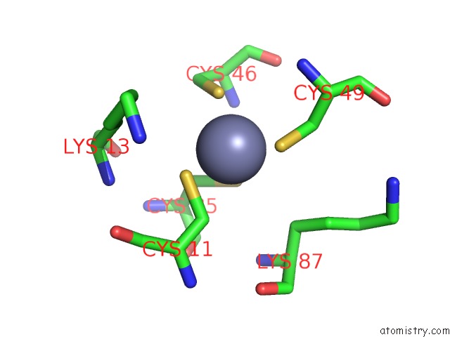
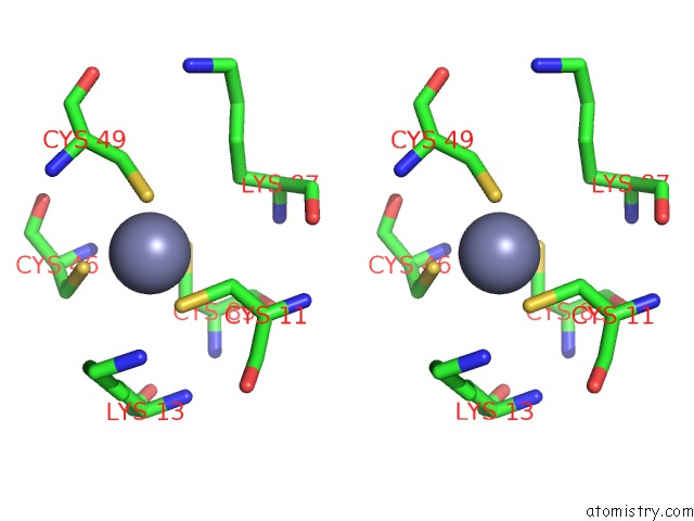
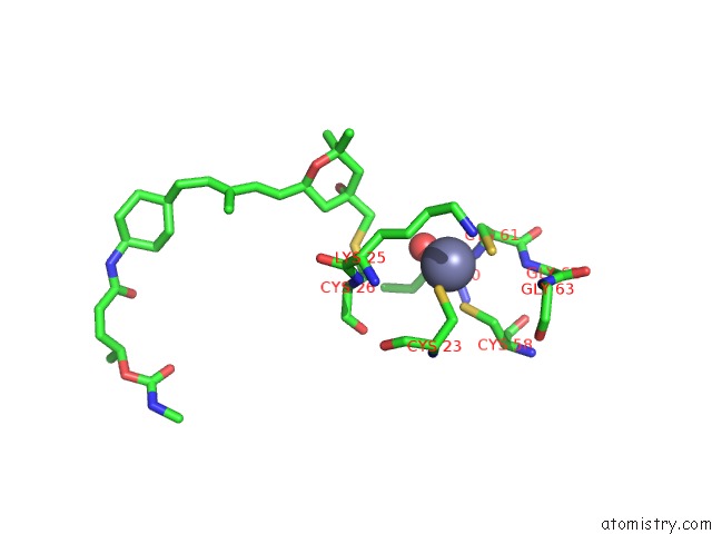
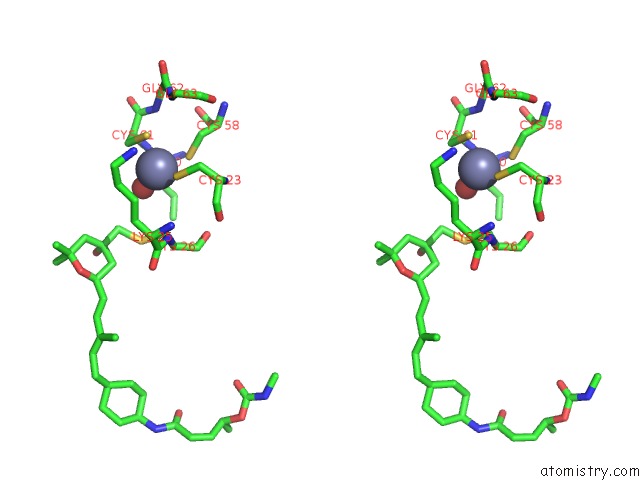
Zinc binding site 1 out of 3 in 7b92
Go back to
Zinc binding site 1 out
of 3 in the Structure of A Minimal SF3B Core in Complex with Sudemycin D6 (Form II)
Mono view
Stereo pair view
Mono view
Stereo pair view
A full contact list of Zinc with other atoms in the Zn binding
site number 1 of Structure of A Minimal SF3B Core in Complex with Sudemycin D6 (Form II) within 5.0Å range:
|
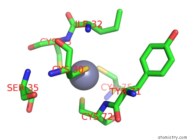
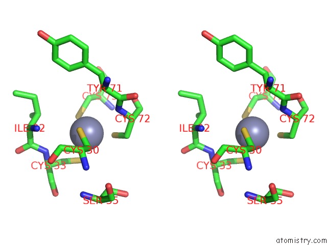
Zinc binding site 2 out of 3 in 7b92
Go back to
Zinc binding site 2 out
of 3 in the Structure of A Minimal SF3B Core in Complex with Sudemycin D6 (Form II)
Mono view
Stereo pair view
Mono view
Stereo pair view
A full contact list of Zinc with other atoms in the Zn binding
site number 2 of Structure of A Minimal SF3B Core in Complex with Sudemycin D6 (Form II) within 5.0Å range:
|
Zinc binding site 3 out of 3 in 7b92
Go back to
Zinc binding site 3 out
of 3 in the Structure of A Minimal SF3B Core in Complex with Sudemycin D6 (Form II)
Mono view
Stereo pair view
Mono view
Stereo pair view
A full contact list of Zinc with other atoms in the Zn binding
site number 3 of Structure of A Minimal SF3B Core in Complex with Sudemycin D6 (Form II) within 5.0Å range:
|
Reference:
C.Cretu,
P.Gee,
X.Liu,
A.Agrawal,
T.V.Nguyen,
A.K.Ghosh,
A.Cook,
M.Jurica,
N.A.Larsen,
V.Pena.
Structural Basis of Intron Selection By U2 Snrnp in the Presence of Covalent Inhibitors. Nat Commun V. 12 4491 2021.
ISSN: ESSN 2041-1723
PubMed: 34301950
DOI: 10.1038/S41467-021-24741-1
Page generated: Tue Oct 29 17:32:04 2024
ISSN: ESSN 2041-1723
PubMed: 34301950
DOI: 10.1038/S41467-021-24741-1
Last articles
Zn in 9J0NZn in 9J0O
Zn in 9J0P
Zn in 9FJX
Zn in 9EKB
Zn in 9C0F
Zn in 9CAH
Zn in 9CH0
Zn in 9CH3
Zn in 9CH1