Zinc in PDB 6nl6: Crystal Structure of Mutant B1 Immunoglobulin-Binding Domain of Streptococcal Protein G (T16F, T18A, V21E, T25L, K28Y, V29I, K31R, Q32H, Y33L, N35K, D36H, N37Q)
Protein crystallography data
The structure of Crystal Structure of Mutant B1 Immunoglobulin-Binding Domain of Streptococcal Protein G (T16F, T18A, V21E, T25L, K28Y, V29I, K31R, Q32H, Y33L, N35K, D36H, N37Q), PDB code: 6nl6
was solved by
T.Huxford,
S.Boguslaw,
B.Maniaci,
with X-Ray Crystallography technique. A brief refinement statistics is given in the table below:
| Resolution Low / High (Å) | 37.38 / 1.40 |
| Space group | P 32 |
| Cell size a, b, c (Å), α, β, γ (°) | 48.292, 48.292, 83.341, 90.00, 90.00, 120.00 |
| R / Rfree (%) | 14.5 / 21.4 |
Other elements in 6nl6:
The structure of Crystal Structure of Mutant B1 Immunoglobulin-Binding Domain of Streptococcal Protein G (T16F, T18A, V21E, T25L, K28Y, V29I, K31R, Q32H, Y33L, N35K, D36H, N37Q) also contains other interesting chemical elements:
| Chlorine | (Cl) | 4 atoms |
Zinc Binding Sites:
The binding sites of Zinc atom in the Crystal Structure of Mutant B1 Immunoglobulin-Binding Domain of Streptococcal Protein G (T16F, T18A, V21E, T25L, K28Y, V29I, K31R, Q32H, Y33L, N35K, D36H, N37Q)
(pdb code 6nl6). This binding sites where shown within
5.0 Angstroms radius around Zinc atom.
In total 6 binding sites of Zinc where determined in the Crystal Structure of Mutant B1 Immunoglobulin-Binding Domain of Streptococcal Protein G (T16F, T18A, V21E, T25L, K28Y, V29I, K31R, Q32H, Y33L, N35K, D36H, N37Q), PDB code: 6nl6:
Jump to Zinc binding site number: 1; 2; 3; 4; 5; 6;
In total 6 binding sites of Zinc where determined in the Crystal Structure of Mutant B1 Immunoglobulin-Binding Domain of Streptococcal Protein G (T16F, T18A, V21E, T25L, K28Y, V29I, K31R, Q32H, Y33L, N35K, D36H, N37Q), PDB code: 6nl6:
Jump to Zinc binding site number: 1; 2; 3; 4; 5; 6;
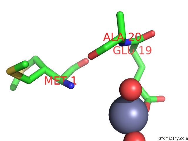
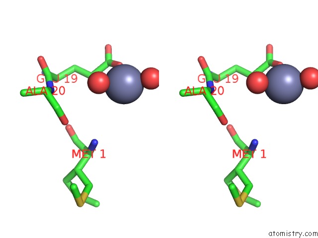
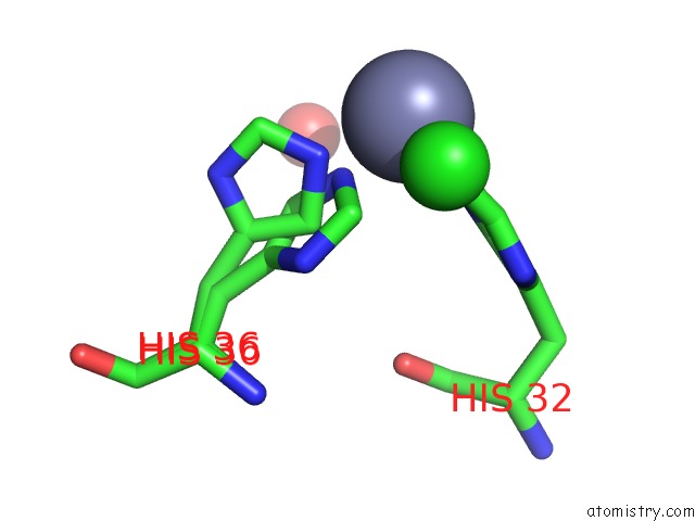
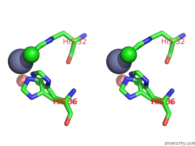
Zinc binding site 1 out of 6 in 6nl6
Go back to
Zinc binding site 1 out
of 6 in the Crystal Structure of Mutant B1 Immunoglobulin-Binding Domain of Streptococcal Protein G (T16F, T18A, V21E, T25L, K28Y, V29I, K31R, Q32H, Y33L, N35K, D36H, N37Q)
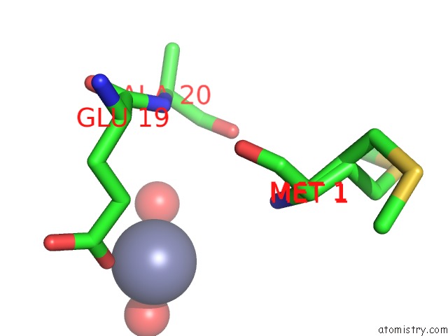
Mono view
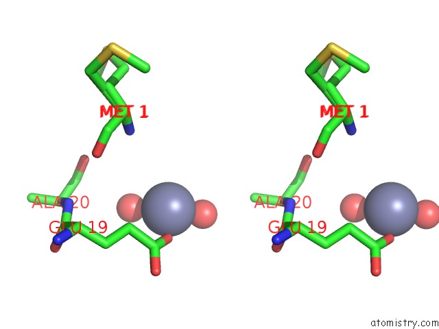
Stereo pair view
Mono view
Stereo pair view
A full contact list of Zinc with other atoms in the Zn binding
site number 1 of Crystal Structure of Mutant B1 Immunoglobulin-Binding Domain of Streptococcal Protein G (T16F, T18A, V21E, T25L, K28Y, V29I, K31R, Q32H, Y33L, N35K, D36H, N37Q) within 5.0Å range:
|
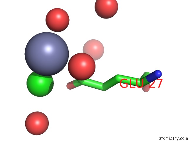
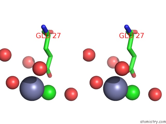
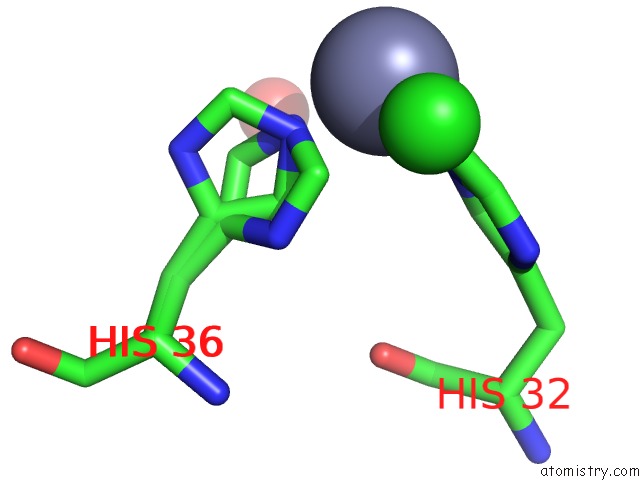
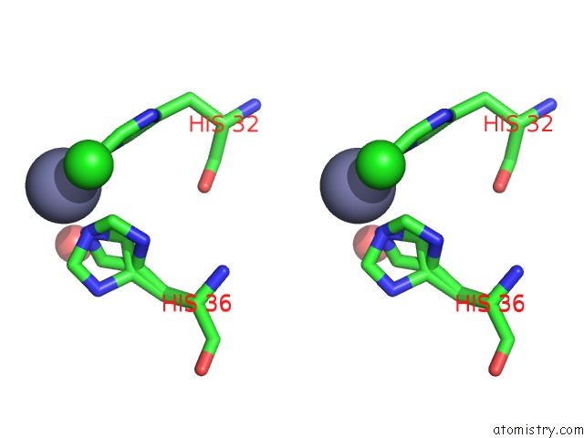
Zinc binding site 2 out of 6 in 6nl6
Go back to
Zinc binding site 2 out
of 6 in the Crystal Structure of Mutant B1 Immunoglobulin-Binding Domain of Streptococcal Protein G (T16F, T18A, V21E, T25L, K28Y, V29I, K31R, Q32H, Y33L, N35K, D36H, N37Q)
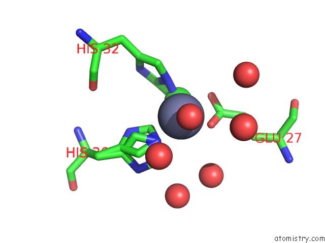
Mono view
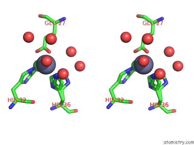
Stereo pair view
Mono view
Stereo pair view
A full contact list of Zinc with other atoms in the Zn binding
site number 2 of Crystal Structure of Mutant B1 Immunoglobulin-Binding Domain of Streptococcal Protein G (T16F, T18A, V21E, T25L, K28Y, V29I, K31R, Q32H, Y33L, N35K, D36H, N37Q) within 5.0Å range:
|
Zinc binding site 3 out of 6 in 6nl6
Go back to
Zinc binding site 3 out
of 6 in the Crystal Structure of Mutant B1 Immunoglobulin-Binding Domain of Streptococcal Protein G (T16F, T18A, V21E, T25L, K28Y, V29I, K31R, Q32H, Y33L, N35K, D36H, N37Q)
Mono view
Stereo pair view
Mono view
Stereo pair view
A full contact list of Zinc with other atoms in the Zn binding
site number 3 of Crystal Structure of Mutant B1 Immunoglobulin-Binding Domain of Streptococcal Protein G (T16F, T18A, V21E, T25L, K28Y, V29I, K31R, Q32H, Y33L, N35K, D36H, N37Q) within 5.0Å range:
|
Zinc binding site 4 out of 6 in 6nl6
Go back to
Zinc binding site 4 out
of 6 in the Crystal Structure of Mutant B1 Immunoglobulin-Binding Domain of Streptococcal Protein G (T16F, T18A, V21E, T25L, K28Y, V29I, K31R, Q32H, Y33L, N35K, D36H, N37Q)
Mono view
Stereo pair view
Mono view
Stereo pair view
A full contact list of Zinc with other atoms in the Zn binding
site number 4 of Crystal Structure of Mutant B1 Immunoglobulin-Binding Domain of Streptococcal Protein G (T16F, T18A, V21E, T25L, K28Y, V29I, K31R, Q32H, Y33L, N35K, D36H, N37Q) within 5.0Å range:
|
Zinc binding site 5 out of 6 in 6nl6
Go back to
Zinc binding site 5 out
of 6 in the Crystal Structure of Mutant B1 Immunoglobulin-Binding Domain of Streptococcal Protein G (T16F, T18A, V21E, T25L, K28Y, V29I, K31R, Q32H, Y33L, N35K, D36H, N37Q)
Mono view
Stereo pair view
Mono view
Stereo pair view
A full contact list of Zinc with other atoms in the Zn binding
site number 5 of Crystal Structure of Mutant B1 Immunoglobulin-Binding Domain of Streptococcal Protein G (T16F, T18A, V21E, T25L, K28Y, V29I, K31R, Q32H, Y33L, N35K, D36H, N37Q) within 5.0Å range:
|
Zinc binding site 6 out of 6 in 6nl6
Go back to
Zinc binding site 6 out
of 6 in the Crystal Structure of Mutant B1 Immunoglobulin-Binding Domain of Streptococcal Protein G (T16F, T18A, V21E, T25L, K28Y, V29I, K31R, Q32H, Y33L, N35K, D36H, N37Q)
Mono view
Stereo pair view
Mono view
Stereo pair view
A full contact list of Zinc with other atoms in the Zn binding
site number 6 of Crystal Structure of Mutant B1 Immunoglobulin-Binding Domain of Streptococcal Protein G (T16F, T18A, V21E, T25L, K28Y, V29I, K31R, Q32H, Y33L, N35K, D36H, N37Q) within 5.0Å range:
|
Reference:
B.Maniaci,
C.H.Lipper,
D.L.Anipindi,
H.Erlandsen,
J.L.Cole,
B.Stec,
T.Huxford,
J.J.Love.
Design of High-Affinity Metal-Controlled Protein Dimers. Biochemistry V. 58 2199 2019.
ISSN: ISSN 0006-2960
PubMed: 30938154
DOI: 10.1021/ACS.BIOCHEM.9B00055
Page generated: Tue Oct 29 03:55:39 2024
ISSN: ISSN 0006-2960
PubMed: 30938154
DOI: 10.1021/ACS.BIOCHEM.9B00055
Last articles
Zn in 9J0NZn in 9J0O
Zn in 9J0P
Zn in 9FJX
Zn in 9EKB
Zn in 9C0F
Zn in 9CAH
Zn in 9CH0
Zn in 9CH3
Zn in 9CH1